 Movie
Movie Controller

[English] 日本語
 Yorodumi
- PDB-2sns: STAPHYLOCOCCAL NUCLEASE. PROPOSED MECHANISM OF ACTION BASED ON ST... -
+ Open data

- Basic information
| Entry | Database: PDB / ID: 2sns | |||||||||
|---|---|---|---|---|---|---|---|---|---|---|
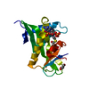
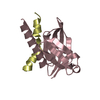
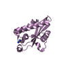
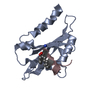
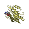
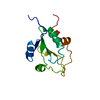
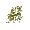
| Title | STAPHYLOCOCCAL NUCLEASE. PROPOSED MECHANISM OF ACTION BASED ON STRUCTURE OF ENZYME-THYMIDINE 3(PRIME),5(PRIME)-BIPHOSPHATE-CALCIUM ION COMPLEX AT 1.5-ANGSTROMS RESOLUTION | |||||||||
 Components Components | THERMONUCLEASE PRECURSOR | |||||||||
 Keywords Keywords | HYDROLASE (PHOSPHORIC DIESTER) | |||||||||
| Function / homology |  Function and homology information Function and homology informationmicrococcal nuclease / 3' overhang single-stranded DNA endonuclease activity / nucleic acid binding / extracellular region / membrane / metal ion binding Similarity search - Function | |||||||||
| Biological species |   Staphylococcus aureus (bacteria) Staphylococcus aureus (bacteria) | |||||||||
| Method |  X-RAY DIFFRACTION / Resolution: 1.5 Å X-RAY DIFFRACTION / Resolution: 1.5 Å | |||||||||
 Authors Authors | Legg, M.J. / Cotton, F.A. / Hazen Jr., E.E. | |||||||||
 Citation Citation | Journal: Thesis, Texas Agricultural and Mechanical University Year: 1977 Title: STAPHYLOCOCCAL NUCLEASE. PROPOSED MECHANISM OF ACTION BASED ON STRUCTURE OF ENZYME-THYMIDINE 3(PRIME),5(PRIME)-BIPHOSPHATE-CALCIUM ION COMPLEX AT 1.5-ANGSTROMS RESOLUTION Authors: Cotton, F.A. / Hazen Jr., E.E. / Legg, M.J. #1: Journal: Proc.Natl.Acad.Sci.USA / Year: 1979 Title: Staphylococcal nuclease: proposed mechanism of action based on structure of enzyme-thymidine 3',5'-bisphosphate-calcium ion complex at 1.5-A resolution. Authors: Cotton, F.A. / Hazenjunior, E.E. / Legg, M.J. #3: Journal: STRUCTURE AND CONFORMATION OF NUCLEIC ACIDS AND PROTEIN-NUCLEIC ACID INTERACTIONS : PROCEEDINGS OF THE FOURTH ANNUAL HARRY STEENBOCK SYMPOSIUM, JUNE 16-19, 1974, MADISON, WISCONSINYear: 1975 Title: The Nucleotide Binding Site of Staphylococcal Nuclease and a New Approach to the Refinement of the Crystal Structures of Biological Macromolecules Authors: Collins, D.M. / Cotton, F.A. / Hazenjunior, E.E. / Legg, M.J. #4: Journal: J.Am.Chem.Soc. / Year: 1974Title: Structure of Bis (Methylguanidinium) Monohydrogen Orthophosphate,A Model for the Arginine-Phosphate Interactions at the Active Site of Staphylococcal Nuclease and Other Phosphohydrolytic Enzymes Authors: Cotton, F.A. / Day, V.W. / Hazenjunior, E.E. / Larsen, S. / Wong, S.T.K. #5: Journal: Cold Spring Harbor Symp.Quant.Biol. / Year: 1972Title: Some Aspects of the Structure of Staphylococcal Nuclease,Part I,Crystallographic Studies Authors: Cotton, F.A. / Bier, C.J. / Day, V.W. / Hazenjunior, E.E. / Larsen, S. #6: Journal: Cold Spring Harbor Symp.Quant.Biol. / Year: 1972Title: Some Aspects of the Structure of Staphylococcal Nuclease,Part II,Studies in Solution Authors: Anfinsen, C.B. / Schechter, A.N. / Taniuchi, H. #7: Journal: J.Biol.Chem. / Year: 1971Title: A High Resolution Structure of an Inhibitor Complex of the Extracellular Nuclease of Staphylococcus Aureus,I.Experimental Procedures and Chain Tracing Authors: Arnone, A. / Bier, C.J. / Cotton, F.A. / Day, V.W. / Hazenjunior, E.E. / Richardson, D.C. / Richardson, J.S. / Yonath, A. #8: Journal: The Enzymes,Third Edition / Year: 1971Title: Staphylococcal Nuclease X-Ray Structure Authors: Cotton, F.A. / Hazenjunior, E.E. | |||||||||
| History |
| |||||||||
| Remark 700 | SHEET THERE ARE TWO BETA SHEETS WHICH FORM A DISTORTED VERSION OF A 5-STRANDED BETA BARREL. THE ...SHEET THERE ARE TWO BETA SHEETS WHICH FORM A DISTORTED VERSION OF A 5-STRANDED BETA BARREL. THE BARREL PATTERN IS -1,-1,+3, +1 IN THE NOTATION OF RICHARDSON. |
- Structure visualization
| Structure viewer | Molecule: MolmilJmol/JSmol |
|---|
- Downloads & links
-Download
| PDBx/mmCIF format | 2sns.cif.gz | 38.3 KB | Display | PDBx/mmCIF format |
|---|---|---|---|---|
| PDB format | pdb2sns.ent.gz | 24.8 KB | Display | PDB format |
| PDBx/mmJSON format | 2sns.json.gz | Tree view | PDBx/mmJSON format | |
| Others |  Other downloads Other downloads |
-Validation report
| Arichive directory | https://data.pdbj.org/pub/pdb/validation_reports/sn/2snsftp://data.pdbj.org/pub/pdb/validation_reports/sn/2sns | HTTPS FTP |
|---|
-Related structure data
| Similar structure data |
|---|


-Links
 PDBj
PDBj- Assembly
| Deposited unit | 
| ||||||||
|---|---|---|---|---|---|---|---|---|---|
| 1 |
| ||||||||
| Unit cell |
| ||||||||
| Atom site foot note | 1: SEE REMARK 6. |
-Components
| #1: Protein | Mass: 16842.346 Da / Num. of mol.: 1 Source method: isolated from a genetically manipulated source Source: (gene. exp.) Staphylococcus aureus (bacteria) / References: UniProt: P00644, micrococcal nuclease |
|---|---|
| #2: Chemical | ChemComp-CA /   Mass: 40.078 Da / Num. of mol.: 1 / Source method: obtained synthetically / Formula: Ca Mass: 40.078 Da / Num. of mol.: 1 / Source method: obtained synthetically / Formula: Ca |
| #3: Chemical | ChemComp-THP /   Type: DNA linking / Mass: 402.188 Da / Num. of mol.: 1 / Source method: obtained synthetically / Formula: C10H16N2O11P2 Type: DNA linking / Mass: 402.188 Da / Num. of mol.: 1 / Source method: obtained synthetically / Formula: C10H16N2O11P2 |
| Compound details | THE ENZYME IS INACTIVE IN THE PRESENCE OF A CALCIUM ION AND THE MOLECULE THYMIDINE 3(PRIME)-5(PRIME) ...THE ENZYME IS INACTIVE IN THE PRESENCE OF A CALCIUM ION AND THE MOLECULE THYMIDINE 3(PRIME)-5(PRIME) DIPHOSPHAT |
| Has protein modification | N |
-Experimental details
-Experiment
| Experiment | Method: X-RAY DIFFRACTION |
|---|
- Sample preparation
| Crystal | Density Matthews: 2.18 Å3/Da / Density % sol: 43.7 % |
|---|---|
| Crystal grow | *PLUS Method: unknown |
-Data collection
| Radiation | Scattering type: x-ray |
|---|---|
| Radiation wavelength | Relative weight: 1 |
| Reflection | *PLUS Highest resolution: 1.5 Å / Num. obs: 18400 / Num. measured all: 25000 |
- Processing
| Refinement | Highest resolution: 1.5 Å Details: THE ELECTRON DENSITY MAP DID NOT DISTINCTLY REVEAL THE LOCATION OF THE FIRST FIVE OR THE LAST EIGHT AMINO ACID RESIDUES (I.E. 1-5 AND 142-149). THE FIRST FIVE RESIDUES COULD BE FITTED TO THE ...Details: THE ELECTRON DENSITY MAP DID NOT DISTINCTLY REVEAL THE LOCATION OF THE FIRST FIVE OR THE LAST EIGHT AMINO ACID RESIDUES (I.E. 1-5 AND 142-149). THE FIRST FIVE RESIDUES COULD BE FITTED TO THE WEAK DENSITY IN SEVERAL DIFFERENT CONFORMATIONS, BUT THERE WAS INSUFFICIENT DENSITY NEAR THE CARBOXY TERMINUS TO PERMIT ANY ATTEMPTS AT FITTING. ATOMIC COORDINATES FOR ALL FITTED ATOMS APPEAR IN THIS ENTRY. | ||||||||||||
|---|---|---|---|---|---|---|---|---|---|---|---|---|---|
| Refinement step | Cycle: LAST / Highest resolution: 1.5 Å
| ||||||||||||
| Refine LS restraints | *PLUS
|
