 Movie
Movie Controller

[English] 日本語
 Yorodumi
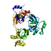
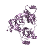
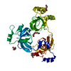
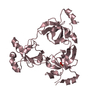
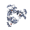
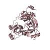
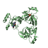
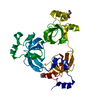
- PDB-2rhy: Crystal structure of the 3-MBT repeats from human L3MBTL1 bound t... -
+ Open data

- Basic information
| Entry | Database: PDB / ID: 2rhy | ||||||
|---|---|---|---|---|---|---|---|
| Title | Crystal structure of the 3-MBT repeats from human L3MBTL1 bound to monomethyl-lysine | ||||||
 Components Components | Lethal(3)malignant brain tumor-like protein | ||||||
 Keywords Keywords | TRANSCRIPTION / Beta barrel / monomethyl-lysine / Alternative splicing / Chromatin regulator / DNA-binding / Metal-binding / Nucleus / Repressor / Transcription regulation / Zinc / Zinc-finger | ||||||
| Function / homology |  Function and homology information Function and homology informationhistone H1 reader activity / SAM domain binding / histone H1K26me1 reader activity / histone H1K26me2 reader activity / constitutive heterochromatin formation / regulation of megakaryocyte differentiation / histone H4K20me2 reader activity / regulation of mitotic nuclear division / hemopoiesis / nucleosome binding ...histone H1 reader activity / SAM domain binding / histone H1K26me1 reader activity / histone H1K26me2 reader activity / constitutive heterochromatin formation / regulation of megakaryocyte differentiation / histone H4K20me2 reader activity / regulation of mitotic nuclear division / hemopoiesis / nucleosome binding / condensed chromosome / Regulation of TP53 Activity through Methylation / heterochromatin formation / chromatin organization / histone binding / regulation of cell cycle / nuclear body / negative regulation of DNA-templated transcription / chromatin binding / chromatin / DNA-templated transcription / zinc ion binding / nucleoplasm / identical protein binding / nucleus Similarity search - Function | ||||||
| Biological species |  Homo sapiens (human) Homo sapiens (human) | ||||||
| Method |  X-RAY DIFFRACTION / Molrep / Resolution: 1.9 Å X-RAY DIFFRACTION / Molrep / Resolution: 1.9 Å | ||||||
 Authors Authors | Li, H. / Patel, D.J. | ||||||
 Citation Citation | Journal: Mol.Cell / Year: 2007 Title: Structural Basis for Lower Lysine Methylation State-Specific Readout by MBT Repeats of L3MBTL1 and an Engineered PHD Finger. Authors: Li, H. / Fischle, W. / Wang, W. / Duncan, E.M. / Liang, L. / Murakami-Ishibe, S. / Allis, C.D. / Patel, D.J. | ||||||
| History |
|
- Structure visualization
| Structure viewer | Molecule: MolmilJmol/JSmol |
|---|
- Downloads & links
-Download
| PDBx/mmCIF format | 2rhy.cif.gz | 89.7 KB | Display | PDBx/mmCIF format |
|---|---|---|---|---|
| PDB format | pdb2rhy.ent.gz | 66.2 KB | Display | PDB format |
| PDBx/mmJSON format | 2rhy.json.gz | Tree view | PDBx/mmJSON format | |
| Others |  Other downloads Other downloads |
-Validation report
| Arichive directory | https://data.pdbj.org/pub/pdb/validation_reports/rh/2rhyftp://data.pdbj.org/pub/pdb/validation_reports/rh/2rhy | HTTPS FTP |
|---|
-Related structure data
| Related structure data |  2rhiC  2rhuC  2rhxC  2rhzC  2ri2C  2ri3C  2ri5C 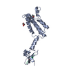 2ri7C  1oz2S C: citing same article ( S: Starting model for refinement |
|---|---|
| Similar structure data |


-Links
 PDBj
PDBj- Assembly
| Deposited unit | 
| ||||||||
|---|---|---|---|---|---|---|---|---|---|
| 1 |
| ||||||||
| Unit cell |
|
-Components
| #1: Protein | Mass: 36693.957 Da / Num. of mol.: 1 / Fragment: Repeates MBT-1, MBT-2, MBT-3; residues 206-519 Source method: isolated from a genetically manipulated source Source: (gene. exp.) Homo sapiens (human) / Gene: L3MBTL, KIAA0681, L3MBT / Plasmid: pGex6p / Production host:  |
|---|---|
| #2: Chemical | ChemComp-MLZ / 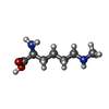  Type: L-peptide linking / Mass: 160.214 Da / Num. of mol.: 1 / Source method: obtained synthetically / Formula: C7H16N2O2 Type: L-peptide linking / Mass: 160.214 Da / Num. of mol.: 1 / Source method: obtained synthetically / Formula: C7H16N2O2 |
| #3: Chemical | ChemComp-PEG /   Mass: 106.120 Da / Num. of mol.: 1 / Source method: obtained synthetically / Formula: C4H10O3 Mass: 106.120 Da / Num. of mol.: 1 / Source method: obtained synthetically / Formula: C4H10O3 |
| #4: Water | ChemComp-HOH /  Mass: 18.015 Da / Num. of mol.: 459 / Source method: isolated from a natural source / Formula: H2O Mass: 18.015 Da / Num. of mol.: 459 / Source method: isolated from a natural source / Formula: H2O |
-Experimental details
-Experiment
| Experiment | Method: X-RAY DIFFRACTION / Number of used crystals: 1 |
|---|
- Sample preparation
| Crystal | Density Matthews: 3.16 Å3/Da / Density % sol: 61.12 % |
|---|---|
| Crystal grow | Temperature: 293 K / Method: vapor diffusion, hanging drop / pH: 6.5 Details: Crystals was generated by pre-incubating the L3MBTL1 (12MG/ML IN 50MM KCL, 25MM TRIS-HCL PH 8.0, 1MM DTT) with a five-fold molar excess of monomethyl-lysine amino acid. Drops were prepared ...Details: Crystals was generated by pre-incubating the L3MBTL1 (12MG/ML IN 50MM KCL, 25MM TRIS-HCL PH 8.0, 1MM DTT) with a five-fold molar excess of monomethyl-lysine amino acid. Drops were prepared by mixing equal volumes of the complex with reservoir solution (7.5% PEG10K, 0.1 M Tris-maleate pH 6.5, 0.1 M ammonium sulfate), VAPOR DIFFUSION, HANGING DROP, temperature 293K |
-Data collection
| Diffraction | Mean temperature: 100 K |
|---|---|
| Diffraction source | Source: ROTATING ANODE / Type: RIGAKU RUH3R / Wavelength: 1.54 Å |
| Detector | Type: RIGAKU RAXIS IV / Detector: IMAGE PLATE / Date: Jun 15, 2007 / Details: mirrors |
| Radiation | Protocol: SINGLE WAVELENGTH / Monochromatic (M) / Laue (L): M / Scattering type: x-ray |
| Radiation wavelength | Wavelength: 1.54 Å / Relative weight: 1 |
| Reflection | Resolution: 1.9→20 Å / Num. all: 37497 / Num. obs: 37422 / % possible obs: 99.8 % / Observed criterion σ(F): -3 / Observed criterion σ(I): -3 / Redundancy: 5.7 % / Biso Wilson estimate: 26.7 Å2 / Rmerge(I) obs: 0.076 / Rsym value: 0.076 / Net I/σ(I): 24.7 |
| Reflection shell | Resolution: 1.9→1.97 Å / Redundancy: 5.5 % / Rmerge(I) obs: 0.485 / Mean I/σ(I) obs: 3.7 / Num. unique all: 3665 / Rsym value: 0.485 / % possible all: 99.3 |
- Processing
| Software |
| |||||||||||||||||||||||||
|---|---|---|---|---|---|---|---|---|---|---|---|---|---|---|---|---|---|---|---|---|---|---|---|---|---|---|
| Refinement | Method to determine structure: Molrep Starting model: 1OZ2 Resolution: 1.9→20 Å / Isotropic thermal model: isotropic / Cross valid method: THROUGHOUT / σ(F): 0 / σ(I): 0 / Stereochemistry target values: Engh & Huber
| |||||||||||||||||||||||||
| Displacement parameters | Biso mean: 32.2 Å2 | |||||||||||||||||||||||||
| Refine analyze |
| |||||||||||||||||||||||||
| Refinement step | Cycle: LAST / Resolution: 1.9→20 Å
| |||||||||||||||||||||||||
| Refine LS restraints |
| |||||||||||||||||||||||||
| LS refinement shell | Resolution: 1.9→1.97 Å
|
