 Movie
Movie Controller

[English] 日本語
 Yorodumi
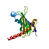
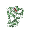
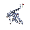
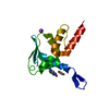
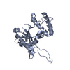
- PDB-2rhe: STRUCTURE OF A NOVEL BENCE-JONES PROTEIN (RHE) FRAGMENT AT 1.6 AN... -
+ Open data

- Basic information
| Entry | Database: PDB / ID: 2rhe | |||||||||
|---|---|---|---|---|---|---|---|---|---|---|
| Title | STRUCTURE OF A NOVEL BENCE-JONES PROTEIN (RHE) FRAGMENT AT 1.6 ANGSTROMS RESOLUTION | |||||||||
 Components Components | BENCE-JONES PROTEIN RHE (LIGHT CHAIN) | |||||||||
 Keywords Keywords | IMMUNOGLOBULIN | |||||||||
| Function / homology | Immunoglobulins / Immunoglobulin-like / Sandwich / Mainly Beta / :  Function and homology information Function and homology information | |||||||||
| Biological species |  Homo sapiens (human) Homo sapiens (human) | |||||||||
| Method |  X-RAY DIFFRACTION / Resolution: 1.6 Å X-RAY DIFFRACTION / Resolution: 1.6 Å | |||||||||
 Authors Authors | Fureyjunior, W. / Wang, B.C. / Yoo, C.S. / Sax, M. | |||||||||
 Citation Citation | Journal: J.Mol.Biol. / Year: 1983 Title: Structure of a novel Bence-Jones protein (Rhe) fragment at 1.6 A resolution. Authors: Furey Jr., W. / Wang, B.C. / Yoo, C.S. / Sax, M. #1: Journal: Acta Crystallogr.,Sect.A / Year: 1979Title: Phase Extension and Refinement of Bence-Jones Protein Rhe (1.9 Angstroms) Authors: Fureyjunior, W. / Wang, B.C. / Yoo, C.S. / Sax, M. #2: Journal: J.Mol.Biol. / Year: 1979Title: Crystal Structure of Bence Jones Protein Rhe (3 Angstroms) and its Unique Domain-Domain Association Authors: Wang, B.-C. / Yoo, C.S. / Sax, M. #3: Journal: J.Mol.Biol. / Year: 1974Title: Structure of a Dimeric Fragment Related to the Lambda-Type Bence-Jones Protein. A Preliminary Study Authors: Wang, B.-C. / Sax, M. | |||||||||
| History |
|
- Structure visualization
| Structure viewer | Molecule: MolmilJmol/JSmol |
|---|
- Downloads & links
-Download
| PDBx/mmCIF format | 2rhe.cif.gz | 36.9 KB | Display | PDBx/mmCIF format |
|---|---|---|---|---|
| PDB format | pdb2rhe.ent.gz | 24.1 KB | Display | PDB format |
| PDBx/mmJSON format | 2rhe.json.gz | Tree view | PDBx/mmJSON format | |
| Others |  Other downloads Other downloads |
-Validation report
| Arichive directory | https://data.pdbj.org/pub/pdb/validation_reports/rh/2rheftp://data.pdbj.org/pub/pdb/validation_reports/rh/2rhe | HTTPS FTP |
|---|
-Related structure data
| Similar structure data |
|---|


-Links
 PDBj
PDBj

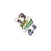
- Assembly
| Deposited unit | 
| ||||||||
|---|---|---|---|---|---|---|---|---|---|
| 1 | 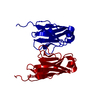
| ||||||||
| Unit cell |
| ||||||||
| Atom site foot note | 1: SEE REMARK 6. / 2: THE SIDE CHAIN OF RESIDUE ILE 35 IS SLIGHTLY DISORDERED. | ||||||||
| Details | THIS MOLECULE EXISTS AS A DIMER BOTH IN SOLUTION AND IN THE CRYSTALS, BUT THE TWO-FOLD AXIS OF DIMERIZATION IS ALIGNED WITH THE CRYSTALLOGRAPHIC Z-AXIS. THUS THE ASYMMETRIC UNIT CONTAINS ONLY A MONOMER. TO GENERATE THE INTACT DIMER, APPLY THE OPERATION -X, -Y, Z TO THE COORDINATES BELOW. |
-Components
| #1: Antibody | Mass: 11840.952 Da / Num. of mol.: 1 Source method: isolated from a genetically manipulated source Source: (gene. exp.) Homo sapiens (human) / Tissue: URINE / References: PIR: S25752 |
|---|---|
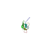
| #2: Water | ChemComp-HOH /  Mass: 18.015 Da / Num. of mol.: 186 / Source method: isolated from a natural source / Formula: H2O Mass: 18.015 Da / Num. of mol.: 186 / Source method: isolated from a natural source / Formula: H2O |
| Has protein modification | Y |
-Experimental details
-Experiment
| Experiment | Method: X-RAY DIFFRACTION |
|---|
- Sample preparation
| Crystal | Density Matthews: 2.57 Å3/Da / Density % sol: 52.05 % | ||||||||||||||||||||||||
|---|---|---|---|---|---|---|---|---|---|---|---|---|---|---|---|---|---|---|---|---|---|---|---|---|---|
| Crystal grow | *PLUS pH: 4.5 / Method: batch method | ||||||||||||||||||||||||
| Components of the solutions | *PLUS
|
-Data collection
| Reflection | *PLUS Highest resolution: 1.6 Å / Num. obs: 12842 / Observed criterion σ(I): 3 / Rmerge F obs: 0.28 / Num. measured all: 16665 |
|---|
- Processing
| Software | Name: PROLSQ / Classification: refinement | ||||||||||||||||||||||||||||||||||||||||||||||||||||||||||||
|---|---|---|---|---|---|---|---|---|---|---|---|---|---|---|---|---|---|---|---|---|---|---|---|---|---|---|---|---|---|---|---|---|---|---|---|---|---|---|---|---|---|---|---|---|---|---|---|---|---|---|---|---|---|---|---|---|---|---|---|---|---|
| Refinement | Resolution: 1.6→10 Å Details: ALTHOUGH THE CHEMICAL EVIDENCE INDICATES THAT RESIDUE 1 IS PCA (PYROLLIDONE CARBOXYLIC ACID) WHICH IS A CYCLIZED GLUTAMIC ACID, THE COORDINATES GIVEN BELOW ARE THOSE OF THE UNCYCLIZED ...Details: ALTHOUGH THE CHEMICAL EVIDENCE INDICATES THAT RESIDUE 1 IS PCA (PYROLLIDONE CARBOXYLIC ACID) WHICH IS A CYCLIZED GLUTAMIC ACID, THE COORDINATES GIVEN BELOW ARE THOSE OF THE UNCYCLIZED GLUTAMIC ACID (GLU) WHICH WAS USED IN THE REFINEMENT PROCESS. ALMOST NO ELECTRON DENSITY WAS OBSERVED FOR THIS RESIDUE IN THE CRYSTALLOGRAPHIC STUDY INDICATING THAT THIS RESIDUE IS BADLY DISORDERED AND DOES NOT PACK WELL INTO THE LATTICE. THEREFORE NO ATTEMPT WAS MADE TO FIT A PCA GROUP TO THE DATA. THIS DISCREPANCY HAS NO EFFECT ON THE REMAINDER OF THE STRUCTURE. THE SOLVENT STRUCTURE HAS BEEN DETERMINED TO THE EXTENT THAT 35 PER CENT OF ALL WATERS WERE FOUND AND REFINED. THE FIRST 102 WATER SITES ARE FULLY OCCUPIED AND ARE BELIEVED TO BE ACCURATELY PLACED. OCCUPANCIES WERE REFINED FOR THE REMAINING WATER SITES, AND THEIR POSITIONS ARE LIKELY TO BE KNOWN WITH LESS ACCURACY. ALL WATER SITES INCLUDED IN THIS ENTRY ARE CHEMICALLY SENSIBLE IN THAT HYDROGEN BONDS ARE FORMED TO SUITABLE DONORS OR ACCEPTORS AND NO BAD PACKING CONTACTS ARE FORMED. WATERS 115, 117, 118, 121 AND 123 ARE PARTICULARLY SIGNIFICANT AND APPEAR TO BE INTEGRAL PARTS OF THE PROTEIN STRUCTURE.
| ||||||||||||||||||||||||||||||||||||||||||||||||||||||||||||
| Refinement step | Cycle: LAST / Resolution: 1.6→10 Å
| ||||||||||||||||||||||||||||||||||||||||||||||||||||||||||||
| Refine LS restraints |
| ||||||||||||||||||||||||||||||||||||||||||||||||||||||||||||
| Refinement | *PLUS Highest resolution: 1.6 Å / Lowest resolution: 10 Å / Num. reflection obs: 12763 / Rfactor obs: 0.149 | ||||||||||||||||||||||||||||||||||||||||||||||||||||||||||||
| Solvent computation | *PLUS | ||||||||||||||||||||||||||||||||||||||||||||||||||||||||||||
| Displacement parameters | *PLUS |
