PROTEIN BINDING / XLF / Cernunnos / non-homologous end joining / DNA Double Strand Break Repair / Alternative splicing / Disease mutation / DNA damage / DNA repair / Nucleus
Function / homology
Function and homology information
DNA ligase IV complex / positive regulation of ligase activity / DNA end binding / DNA-dependent protein kinase-DNA ligase 4 complex / immunoglobulin V(D)J recombination / nonhomologous end joining complex / response to ionizing radiation / T cell differentiation / B cell differentiation / DNA polymerase binding ...DNA ligase IV complex / positive regulation of ligase activity / DNA end binding / DNA-dependent protein kinase-DNA ligase 4 complex / immunoglobulin V(D)J recombination / nonhomologous end joining complex / response to ionizing radiation / T cell differentiation / B cell differentiation / DNA polymerase binding / telomere maintenance / central nervous system development / Nonhomologous End-Joining (NHEJ) / double-strand break repair via nonhomologous end joining / fibrillar center / site of double-strand break / DNA binding / nucleoplasm / nucleus Similarity search - Function
Helix hairpin bin / DNA double-strand break repair and VJ recombination XRCC4, N-terminal / Dna Repair Protein Xrcc4; Chain: A, domain 1 / XLF, N-terminal / : / : / XLF N-terminal domain / XLF protein coiled-coil region / XRCC4-like, N-terminal domain superfamily / Beta Complex ...Helix hairpin bin / DNA double-strand break repair and VJ recombination XRCC4, N-terminal / Dna Repair Protein Xrcc4; Chain: A, domain 1 / XLF, N-terminal / : / : / XLF N-terminal domain / XLF protein coiled-coil region / XRCC4-like, N-terminal domain superfamily / Beta Complex / Helix Hairpins / Orthogonal Bundle / Mainly Beta / Mainly Alpha Similarity search - Domain/homology
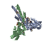
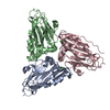
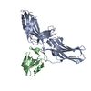
The dimer present within the asymmetric unit is believed to represent the biological assembly. This protein elutes from a gel filtration column with an apparent molecular mass equivalent to a dimer of XLF.
-
Components
#1: Protein
Non-homologousend-joiningfactor1 / Protein cernunnos / XRCC4-like factor
Mass: 26708.400 Da / Num. of mol.: 2 / Fragment: N-terminal fragment, unp residues 1-224 Source method: isolated from a genetically manipulated source Source: (gene. exp.) Homo sapiens (human) / Gene: NHEJ1, XLF / Production host: Escherichia coli (E. coli) / Strain (production host): Rossetta(DE3) / References: UniProt: Q9H9Q4
Resolution: 2.5→2.59 Å / Rmerge(I) obs: 0.43 / Mean I/σ(I) obs: 3.7 / % possible all: 99.9
-
Processing
Software
Name
Version
Classification
REFMAC
5.2.0019
refinement
CBASS
datacollection
CrystalClear
datareduction
CrystalClear
datascaling
CNS
phasing
Refinement
Method to determine structure: SAD / Resolution: 2.5→25 Å / Cor.coef. Fo:Fc: 0.886 / Cor.coef. Fo:Fc free: 0.859 / SU B: 18.735 / SU ML: 0.223 / Cross valid method: THROUGHOUT / σ(F): 3 / ESU R Free: 0.379 / Stereochemistry target values: MAXIMUM LIKELIHOOD / Details: HYDROGENS HAVE BEEN ADDED IN THE RIDING POSITIONS
Rfactor
Num. reflection
% reflection
Selection details
Rfree
0.28674
1031
6.8 %
RANDOM
Rwork
0.25083
-
-
-
all
0.2952
18163
-
-
obs
0.25329
14110
100 %
-
Solvent computation
Ion probe radii: 0.8 Å / Shrinkage radii: 0.8 Å / VDW probe radii: 1.4 Å / Solvent model: BABINET MODEL WITH MASK
Displacement parameters
Biso mean: 45.424 Å2
Baniso -1
Baniso -2
Baniso -3
1-
-1.31 Å2
0 Å2
0 Å2
2-
-
2.24 Å2
0 Å2
3-
-
-
-0.93 Å2
Refinement step
Cycle: LAST / Resolution: 2.5→25 Å
Protein
Nucleic acid
Ligand
Solvent
Total
Num. atoms
3462
0
0
282
3744
Refine LS restraints
Refine-ID
Type
Dev ideal
Dev ideal target
Number
X-RAY DIFFRACTION
r_bond_refined_d
0.016
0.022
3537
X-RAY DIFFRACTION
r_angle_refined_deg
1.616
1.969
4795
X-RAY DIFFRACTION
r_dihedral_angle_1_deg
6.315
5
429
X-RAY DIFFRACTION
r_dihedral_angle_2_deg
39.123
24.847
163
X-RAY DIFFRACTION
r_dihedral_angle_3_deg
18.213
15
635
X-RAY DIFFRACTION
r_dihedral_angle_4_deg
11.609
15
17
X-RAY DIFFRACTION
r_chiral_restr
0.112
0.2
548
X-RAY DIFFRACTION
r_gen_planes_refined
0.006
0.02
2635
X-RAY DIFFRACTION
r_nbd_refined
0.265
0.2
1779
X-RAY DIFFRACTION
r_nbtor_refined
0.31
0.2
2410
X-RAY DIFFRACTION
r_xyhbond_nbd_refined
0.208
0.2
229
X-RAY DIFFRACTION
r_symmetry_vdw_refined
0.302
0.2
33
X-RAY DIFFRACTION
r_symmetry_hbond_refined
0.225
0.2
5
X-RAY DIFFRACTION
r_mcbond_it
1.53
1.5
2229
X-RAY DIFFRACTION
r_mcangle_it
1.491
2
3493
X-RAY DIFFRACTION
r_scbond_it
2.642
3
1475
X-RAY DIFFRACTION
r_scangle_it
3.951
4.5
1302
LS refinement shell
Resolution: 2.5→2.564 Å / Total num. of bins used: 20
Rfactor
Num. reflection
% reflection
Rfree
0.3
65
-
Rwork
0.224
1073
-
obs
-
-
100 %
+
About Yorodumi
-
News
-
Feb 9, 2022. New format data for meta-information of EMDB entries
New format data for meta-information of EMDB entries
Version 3 of the EMDB header file is now the official format.
The previous official version 1.9 will be removed from the archive.
In the structure databanks used in Yorodumi, some data are registered as the other names, "COVID-19 virus" and "2019-nCoV". Here are the details of the virus and the list of structure data.
Jan 31, 2019. EMDB accession codes are about to change! (news from PDBe EMDB page)
EMDB accession codes are about to change! (news from PDBe EMDB page)
The allocation of 4 digits for EMDB accession codes will soon come to an end. Whilst these codes will remain in use, new EMDB accession codes will include an additional digit and will expand incrementally as the available range of codes is exhausted. The current 4-digit format prefixed with “EMD-” (i.e. EMD-XXXX) will advance to a 5-digit format (i.e. EMD-XXXXX), and so on. It is currently estimated that the 4-digit codes will be depleted around Spring 2019, at which point the 5-digit format will come into force.
The EM Navigator/Yorodumi systems omit the EMD- prefix.
Related info.:Q: What is EMD? / ID/Accession-code notation in Yorodumi/EM Navigator
Yorodumi is a browser for structure data from EMDB, PDB, SASBDB, etc.
This page is also the successor to EM Navigator detail page, and also detail information page/front-end page for Omokage search.
The word "yorodu" (or yorozu) is an old Japanese word meaning "ten thousand". "mi" (miru) is to see.
Related info.:EMDB / PDB / SASBDB / Comparison of 3 databanks / Yorodumi Search / Aug 31, 2016. New EM Navigator & Yorodumi / Yorodumi Papers / Jmol/JSmol / Function and homology information / Changes in new EM Navigator and Yorodumi
 Movie
Movie Controller


 Open data
 Basic information
 Components
 Keywords
 Function and homology information
 Homo sapiens (human)
 X-RAY DIFFRACTION /
  Authors
 Citation
 Structure visualization
 Downloads & links
 Other downloads


 PDBj
PDBj

 Assembly

 Mass: 18.015 Da / Num. of mol.: 282 / Source method: isolated from a natural source / Formula: H2O
 Sample preparation
 / Beamline: X8C / Wavelength: 0.979 Å
 Processing