Hydrolases; Acting on ester bonds; Phosphoric-diester hydrolases / 3',5'-cyclic-nucleotide phosphodiesterase activity / signal transduction / metal ion binding Similarity search - Function
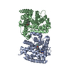
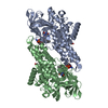
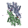
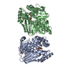
Authors state that there are two molecules of LmjPDEB1 in the crystallographic asymmetric unit. They form a dimer in the crystal. However, it is not clear if the dimer is biologically relevant.
-
Components
#1: Protein
ClassIphosphodiesterasePDEB1
Mass: 40563.922 Da / Num. of mol.: 2 / Fragment: catalytic domain Source method: isolated from a genetically manipulated source Source: (gene. exp.) Leishmania major (eukaryote) / Gene: PDEB1 / Plasmid: pET28a / Species (production host): Escherichia coli / Production host: Escherichia coli BL21 (bacteria) / Strain (production host): BL21 References: UniProt: Q6S996, 3',5'-cyclic-nucleotide phosphodiesterase
Mass: 18.015 Da / Num. of mol.: 501 / Source method: isolated from a natural source / Formula: H2O
-
Experimental details
-
Experiment
Experiment
Method: X-RAY DIFFRACTION / Number of used crystals: 1
-
Sample preparation
Crystal
Density Matthews: 2.18 Å3/Da / Density % sol: 43.71 %
Crystal grow
Temperature: 277 K / Method: vapor diffusion, hanging drop / pH: 7.5 Details: The protein drop was prepared by mixing 2 microliter of the LmjPDEB1-IBMX complex with 2 microliter well buffer of 30% PEG3350, 0.1 M HEPES, pH 7.5, VAPOR DIFFUSION, HANGING DROP, temperature 277K
In the structure databanks used in Yorodumi, some data are registered as the other names, "COVID-19 virus" and "2019-nCoV". Here are the details of the virus and the list of structure data.
Jan 31, 2019. EMDB accession codes are about to change! (news from PDBe EMDB page)
EMDB accession codes are about to change! (news from PDBe EMDB page)
The allocation of 4 digits for EMDB accession codes will soon come to an end. Whilst these codes will remain in use, new EMDB accession codes will include an additional digit and will expand incrementally as the available range of codes is exhausted. The current 4-digit format prefixed with “EMD-” (i.e. EMD-XXXX) will advance to a 5-digit format (i.e. EMD-XXXXX), and so on. It is currently estimated that the 4-digit codes will be depleted around Spring 2019, at which point the 5-digit format will come into force.
The EM Navigator/Yorodumi systems omit the EMD- prefix.
Related info.:Q: What is EMD? / ID/Accession-code notation in Yorodumi/EM Navigator
Yorodumi is a browser for structure data from EMDB, PDB, SASBDB, etc.
This page is also the successor to EM Navigator detail page, and also detail information page/front-end page for Omokage search.
The word "yorodu" (or yorozu) is an old Japanese word meaning "ten thousand". "mi" (miru) is to see.
Related info.:EMDB / PDB / SASBDB / Comparison of 3 databanks / Yorodumi Search / Aug 31, 2016. New EM Navigator & Yorodumi / Yorodumi Papers / Jmol/JSmol / Function and homology information / Changes in new EM Navigator and Yorodumi
 Movie
Movie Controller


 Open data
 Basic information
 Components
 Keywords
 Function and homology information
 Leishmania major (eukaryote)
 X-RAY DIFFRACTION /
  Authors
 Citation
 Structure visualization
 Downloads & links
 Other downloads
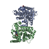


 PDBj
PDBj

 Assembly


 Mass: 65.409 Da / Num. of mol.: 2 / Source method: obtained synthetically / Formula: Zn
 Mass: 24.305 Da / Num. of mol.: 2 / Source method: obtained synthetically / Formula: Mg
 Mass: 222.244 Da / Num. of mol.: 2 / Source method: obtained synthetically / Formula: C10H14N4O2
 Mass: 18.015 Da / Num. of mol.: 501 / Source method: isolated from a natural source / Formula: H2O
 Sample preparation
 / Beamline: X29A / Wavelength: 1.08 Å
 Processing