 Movie
Movie Controller

[English] 日本語
 Yorodumi
- PDB-2r6v: Crystal structure of FMN-binding protein (NP_142786.1) from Pyroc... -
+ Open data

- Basic information
| Entry | Database: PDB / ID: 2r6v | ||||||
|---|---|---|---|---|---|---|---|
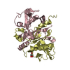
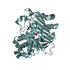
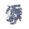
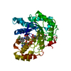
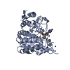
| Title | Crystal structure of FMN-binding protein (NP_142786.1) from Pyrococcus horikoshii at 1.35 A resolution | ||||||
 Components Components | Uncharacterized protein PH0856 | ||||||
 Keywords Keywords | FLAVOPROTEIN / NP_142786.1 / FMN-binding protein / Flavin reductase like domain / Structural Genomics / Joint Center for Structural Genomics / JCSG / Protein Structure Initiative / PSI-2 | ||||||
| Function / homology |  Function and homology information Function and homology information | ||||||
| Biological species |   Pyrococcus horikoshii (archaea) Pyrococcus horikoshii (archaea) | ||||||
| Method |  X-RAY DIFFRACTION / SYNCHROTRON / MAD / Resolution: 1.25 Å X-RAY DIFFRACTION / SYNCHROTRON / MAD / Resolution: 1.25 Å | ||||||
 Authors Authors | Joint Center for Structural Genomics (JCSG) | ||||||
 Citation Citation | Journal: To be published Title: Crystal structure of FMN-binding protein (NP_142786.1) from Pyrococcus horikoshii at 1.35 A resolution Authors: Joint Center for Structural Genomics (JCSG) | ||||||
| History |
| ||||||
| Remark 999 | SEQUENCE THE CONSTRUCT WAS EXPRESSED WITH A PURIFICATION TAG MGSDKIHHHHHHENLYFQG. |
- Structure visualization
| Structure viewer | Molecule: MolmilJmol/JSmol |
|---|
- Downloads & links
-Download
| PDBx/mmCIF format | 2r6v.cif.gz | 107.8 KB | Display | PDBx/mmCIF format |
|---|---|---|---|---|
| PDB format | pdb2r6v.ent.gz | 82.3 KB | Display | PDB format |
| PDBx/mmJSON format | 2r6v.json.gz | Tree view | PDBx/mmJSON format | |
| Others |  Other downloads Other downloads |
-Validation report
| Arichive directory | https://data.pdbj.org/pub/pdb/validation_reports/r6/2r6vftp://data.pdbj.org/pub/pdb/validation_reports/r6/2r6v | HTTPS FTP |
|---|
-Related structure data
| Similar structure data | |
|---|---|
| Other databases |


-Links
 PDBj
PDBj- Assembly
| Deposited unit | 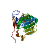
| |||||||||
|---|---|---|---|---|---|---|---|---|---|---|
| 1 | 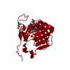
| |||||||||
| Unit cell |
| |||||||||
| Components on special symmetry positions |
|
-Components
| #1: Protein | Mass: 21925.521 Da / Num. of mol.: 1 Source method: isolated from a genetically manipulated source Source: (gene. exp.) Pyrococcus horikoshii (archaea) / Strain: OT3 / Gene: NP_142786.1, PH0856 / Plasmid: speedET / Production host:  |
|---|---|
| #2: Chemical | ChemComp-FMN /   Mass: 456.344 Da / Num. of mol.: 1 / Source method: obtained synthetically / Formula: C17H21N4O9P Mass: 456.344 Da / Num. of mol.: 1 / Source method: obtained synthetically / Formula: C17H21N4O9P |
| #3: Chemical | ChemComp-NCA /   Mass: 122.125 Da / Num. of mol.: 1 / Source method: obtained synthetically / Formula: C6H6N2O / Comment: medication*YM Mass: 122.125 Da / Num. of mol.: 1 / Source method: obtained synthetically / Formula: C6H6N2O / Comment: medication*YM |
| #4: Chemical | ChemComp-EDO /   Mass: 62.068 Da / Num. of mol.: 1 / Source method: obtained synthetically / Formula: C2H6O2 Mass: 62.068 Da / Num. of mol.: 1 / Source method: obtained synthetically / Formula: C2H6O2 |
| #5: Water | ChemComp-HOH /  Mass: 18.015 Da / Num. of mol.: 208 / Source method: isolated from a natural source / Formula: H2O Mass: 18.015 Da / Num. of mol.: 208 / Source method: isolated from a natural source / Formula: H2O |
| Has protein modification | Y |
-Experimental details
-Experiment
| Experiment | Method: X-RAY DIFFRACTION / Number of used crystals: 1 |
|---|
- Sample preparation
| Crystal | Density Matthews: 1.88 Å3/Da / Density % sol: 34.57 % Description: THE HIGH RESOLUTION DATA FROM THE PEAK WAVELENGTH IS INCOMPLETE. LOWER RESOLUTION DATA FROM THE INFLECTION AND HIGH ENERGY REMOTE WAVELENGTHS WERE MERGED WITH THE PEAK DATA TO INCREASE ...Description: THE HIGH RESOLUTION DATA FROM THE PEAK WAVELENGTH IS INCOMPLETE. LOWER RESOLUTION DATA FROM THE INFLECTION AND HIGH ENERGY REMOTE WAVELENGTHS WERE MERGED WITH THE PEAK DATA TO INCREASE THE COMPLETENESS TO 1.4 A. THIS DATA WAS USED FOR REFINEMENT AND THE STATISTICS ABOVE ARE FROM THIS MERGED DATA SET. |
|---|---|
| Crystal grow | Temperature: 277 K / Method: vapor diffusion, sitting drop / pH: 6.6 Details: NANODROP, 0.2M Ammonium formate, 20.0% PEG 3350, No Buffer pH 6.6, VAPOR DIFFUSION, SITTING DROP, temperature 277K |
-Data collection
| Diffraction | Mean temperature: 100 K | |||||||||||||||||||||||||||||||||||||||||||||||||||||||||||||||||||||||||||||
|---|---|---|---|---|---|---|---|---|---|---|---|---|---|---|---|---|---|---|---|---|---|---|---|---|---|---|---|---|---|---|---|---|---|---|---|---|---|---|---|---|---|---|---|---|---|---|---|---|---|---|---|---|---|---|---|---|---|---|---|---|---|---|---|---|---|---|---|---|---|---|---|---|---|---|---|---|---|---|
| Diffraction source | Source: SYNCHROTRON / Site: SSRL  / Beamline: BL11-1 / Wavelength: 0.91837, 0.97939, 0.97910 / Beamline: BL11-1 / Wavelength: 0.91837, 0.97939, 0.97910 | |||||||||||||||||||||||||||||||||||||||||||||||||||||||||||||||||||||||||||||
| Detector | Type: MARMOSAIC 325 mm CCD / Detector: CCD / Date: Jul 20, 2007 / Details: Flat mirror (vertical focusing) | |||||||||||||||||||||||||||||||||||||||||||||||||||||||||||||||||||||||||||||
| Radiation | Monochromator: Single crystal Si(111) bent (horizontal focusing) Protocol: MAD / Monochromatic (M) / Laue (L): M / Scattering type: x-ray | |||||||||||||||||||||||||||||||||||||||||||||||||||||||||||||||||||||||||||||
| Radiation wavelength |
| |||||||||||||||||||||||||||||||||||||||||||||||||||||||||||||||||||||||||||||
| Reflection | Resolution: 1.25→44.588 Å / Num. obs: 41184 / % possible obs: 84.5 % / Redundancy: 18.3 % / Biso Wilson estimate: 15.957 Å2 / Rmerge(I) obs: 0.068 / Net I/σ(I): 23.93 | |||||||||||||||||||||||||||||||||||||||||||||||||||||||||||||||||||||||||||||
| Reflection shell | Diffraction-ID: 1
|
-Phasing
| Phasing | Method: MAD |
|---|
- Processing
| Software |
| ||||||||||||||||||||||||||||||||||||||||||||||||||||||||||||||||||||||||||||||||||||||||||||||||||||||||||||||||||||||||||||||||||||||||||||
|---|---|---|---|---|---|---|---|---|---|---|---|---|---|---|---|---|---|---|---|---|---|---|---|---|---|---|---|---|---|---|---|---|---|---|---|---|---|---|---|---|---|---|---|---|---|---|---|---|---|---|---|---|---|---|---|---|---|---|---|---|---|---|---|---|---|---|---|---|---|---|---|---|---|---|---|---|---|---|---|---|---|---|---|---|---|---|---|---|---|---|---|---|---|---|---|---|---|---|---|---|---|---|---|---|---|---|---|---|---|---|---|---|---|---|---|---|---|---|---|---|---|---|---|---|---|---|---|---|---|---|---|---|---|---|---|---|---|---|---|---|---|
| Refinement | Method to determine structure: MAD / Resolution: 1.25→44.588 Å / Cor.coef. Fo:Fc: 0.978 / Cor.coef. Fo:Fc free: 0.971 / SU B: 1.236 / SU ML: 0.024 / Cross valid method: THROUGHOUT / σ(F): 0 / ESU R: 0.05 / ESU R Free: 0.045 Stereochemistry target values: MAXIMUM LIKELIHOOD WITH PHASES Details: 1. HYDROGENS HAVE BEEN ADDED IN THE RIDING POSITIONS. 2. A MET-INHIBITION PROTOCOL WAS USED FOR SELENOMETHIONINE INCORPORATION DURING PROTEIN EXPRESSION. THE OCCUPANCY OF THE SE ATOMS IN THE ...Details: 1. HYDROGENS HAVE BEEN ADDED IN THE RIDING POSITIONS. 2. A MET-INHIBITION PROTOCOL WAS USED FOR SELENOMETHIONINE INCORPORATION DURING PROTEIN EXPRESSION. THE OCCUPANCY OF THE SE ATOMS IN THE MSE RESIDUES WAS REDUCED TO 0.80 FOR THE REDUCED SCATTERING POWER DUE TO PARTIAL S-MET INCORPORATION. 3. EDO IS FROM CRYO CONDITION. FMN IS ASSIGNED BASED ON DENSITY, STRUCTURAL HOMOLOGS AS WELL AS INTERACTION WITH PROTEIN. NCA (NICOTINAMIDE) IS TENTATIVELY ASSIGNED BASED ON DENSITY AND INTERACTION. NCA COULD BE A PORTION OF THE PUTATIVE LIGAND NADP+ (PDB 1I0S) OR OTHER RELATED COMPOUNDS. 4. DUE TO LOW COMPLETENESS OF THE HIGH RESOLUTION DATA FROM THE PEAK WAVELENGTH, REFINEMENT WAS AGAINST DATA MERGED FROM ALL THREE WAVELENGTHS. THIS MERGED DATA IS 85 % COMPLETE OVERALL. THE NOMINAL RESOLUTION IS 1.35A WITH 4292 OBSERVED REFLECTIONS BETWEEN 1.35-1.25A (44 % COMPLETE FOR THIS SHELL) INCLUDED IN THE REFINEMENT.
| ||||||||||||||||||||||||||||||||||||||||||||||||||||||||||||||||||||||||||||||||||||||||||||||||||||||||||||||||||||||||||||||||||||||||||||
| Solvent computation | Ion probe radii: 0.8 Å / Shrinkage radii: 0.8 Å / VDW probe radii: 1.2 Å / Solvent model: BABINET MODEL WITH MASK | ||||||||||||||||||||||||||||||||||||||||||||||||||||||||||||||||||||||||||||||||||||||||||||||||||||||||||||||||||||||||||||||||||||||||||||
| Displacement parameters | Biso mean: 12.524 Å2
| ||||||||||||||||||||||||||||||||||||||||||||||||||||||||||||||||||||||||||||||||||||||||||||||||||||||||||||||||||||||||||||||||||||||||||||
| Refinement step | Cycle: LAST / Resolution: 1.25→44.588 Å
| ||||||||||||||||||||||||||||||||||||||||||||||||||||||||||||||||||||||||||||||||||||||||||||||||||||||||||||||||||||||||||||||||||||||||||||
| Refine LS restraints |
| ||||||||||||||||||||||||||||||||||||||||||||||||||||||||||||||||||||||||||||||||||||||||||||||||||||||||||||||||||||||||||||||||||||||||||||
| LS refinement shell | Resolution: 1.25→1.283 Å / Total num. of bins used: 20
|
