[English] 日本語
Yorodumi
- PDB-2r16: Crystal Structure of bovine neurexin 1 alpha LNS/LG domain 4 (wit... -

+
Open data


ID or keywords:

Loading...

-
Basic information

Entry
Database: PDB / ID: 2r16
Title
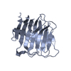
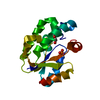
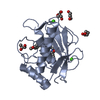
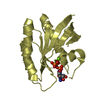
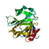
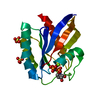
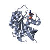
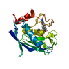
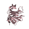
Crystal Structure of bovine neurexin 1 alpha LNS/LG domain 4 (with no splice insert)
ComponentsNeurexin-1-alpha
KeywordsCELL ADHESION / SPLICING / beta-sandwich
Function / homology
Function and homology information


neuroligin family protein binding / cell projection / presynaptic membrane / cell adhesion / metal ion binding
Similarity search - Function
Neurexin/syndecan/glycophorin C / putative band 4.1 homologues' binding motif / Laminin G domain / Laminin G domain profile. / Laminin G domain / Laminin G domain / EGF-like domain / EGF-type aspartate/asparagine hydroxylation site / Aspartic acid and asparagine hydroxylation site. / Epidermal growth factor-like domain. ...Neurexin/syndecan/glycophorin C / putative band 4.1 homologues' binding motif / Laminin G domain / Laminin G domain profile. / Laminin G domain / Laminin G domain / EGF-like domain / EGF-type aspartate/asparagine hydroxylation site / Aspartic acid and asparagine hydroxylation site. / Epidermal growth factor-like domain. / Jelly Rolls - #200 / EGF-like domain profile. / EGF-like domain / Concanavalin A-like lectin/glucanase domain superfamily / Jelly Rolls / Sandwich / Mainly Beta
Similarity search - Domain/homology
Biological speciesBos taurus (cattle)
MethodX-RAY DIFFRACTION / SYNCHROTRON / MOLECULAR REPLACEMENT / Resolution: 1.04 Å
AuthorsRudenko, G.
CitationJournal: Structure / Year: 2008
Title: Regulation of Neurexin 1beta Tertiary Structure and Ligand Binding through Alternative Splicing
Authors: Shen, K.C. / Kuczynska, D.A. / Wu, I.J. / Murray, B.H. / Sheckler, L.R. / Rudenko, G.
History
DepositionAug 22, 2007Deposition site: RCSB / Processing site: RCSB
Revision 1.0Mar 25, 2008Provider: repository / Type: Initial release
Revision 1.1Jul 13, 2011Group: Version format compliance
Revision 1.2Oct 25, 2017Group: Refinement description / Category: software / Item: _software.name
Revision 1.3Jul 24, 2019Group: Data collection / Refinement description / Category: software
Item: _software.classification / _software.name / _software.version
Revision 1.4Aug 30, 2023Group: Data collection / Database references ...Data collection / Database references / Derived calculations / Refinement description
Category: chem_comp_atom / chem_comp_bond ...chem_comp_atom / chem_comp_bond / database_2 / pdbx_initial_refinement_model / struct_ref_seq_dif / struct_site
Item: _database_2.pdbx_DOI / _database_2.pdbx_database_accession ..._database_2.pdbx_DOI / _database_2.pdbx_database_accession / _struct_ref_seq_dif.details / _struct_site.pdbx_auth_asym_id / _struct_site.pdbx_auth_comp_id / _struct_site.pdbx_auth_seq_id

-
Structure visualization

Structure viewerMolecule:
MolmilJmol/JSmol

Downloads & links

-
Assembly

Deposited unit
A: Neurexin-1-alpha
hetero molecules


Theoretical massNumber of molelcules
Total (without water)20,5792
Polymers20,5391
Non-polymers401
Water2,342130
1


  • Idetical with deposited unit
  • defined by author&software
TypeNameSymmetry operationNumber
identity operation1_555x,y,z1
MethodPISA
Unit cell
Length a, b, c (Å)90.322, 90.322, 39.624
Angle α, β, γ (deg.)90.00, 90.00, 120.00
Int Tables number173
Space group name H-MP63

-
Components

#1: Protein Neurexin-1-alpha / neurexin I-alpha


Mass: 20539.264 Da / Num. of mol.: 1
Source method: isolated from a genetically manipulated source
Source: (gene. exp.) Bos taurus (cattle) / Gene: NRXN1 / Plasmid: pGEX-KG / Production host: Escherichia coli (E. coli) / Strain (production host): Origami B / References: UniProt: Q28146
#2: Chemical ChemComp-CA / CALCIUM ION


Mass: 40.078 Da / Num. of mol.: 1 / Source method: obtained synthetically / Formula: Ca
#3: Water ChemComp-HOH / water


Mass: 18.015 Da / Num. of mol.: 130 / Source method: isolated from a natural source / Formula: H2O

-
Experimental details

-
Experiment

ExperimentMethod: X-RAY DIFFRACTION / Number of used crystals: 1

-
Sample preparation

CrystalDensity Matthews: 2.27 Å3/Da / Density % sol: 45.85 %
Crystal growTemperature: 293 K / Method: vapor diffusion, hanging drop / pH: 9
Details: 12.5% isopropanol, 0.1 M CHES pH 9.0, 5 mM CaCl2, , VAPOR DIFFUSION, HANGING DROP, temperature 293K

-
Data collection

DiffractionMean temperature: 103 K
Diffraction sourceSource: SYNCHROTRON / Site: APS / Beamline: 23-ID-B / Wavelength: 0.96112 Å
DetectorType: MARMOSAIC 300 mm CCD / Detector: CCD / Date: Aug 16, 2006
RadiationMonochromator: see beam-line specifications / Protocol: SINGLE WAVELENGTH / Monochromatic (M) / Laue (L): M / Scattering type: x-ray
Radiation wavelengthWavelength: 0.96112 Å / Relative weight: 1
ReflectionResolution: 1.04→23.7 Å / Num. all: 82197 / Num. obs: 82197 / % possible obs: 92.9 % / Observed criterion σ(F): 0 / Observed criterion σ(I): 0 / Redundancy: 12.6 % / Biso Wilson estimate: 7.5 Å2 / Rmerge(I) obs: 0.07 / Net I/σ(I): 37.9
Reflection shellResolution: 1.04→1.08 Å / Redundancy: 7.6 % / Rmerge(I) obs: 0.296 / Mean I/σ(I) obs: 6.2 / Num. unique all: 5319 / Rsym value: 0.296 / % possible all: 60.5

-
Processing

Software
NameVersionClassification
REFMAC5.3refinement
CNSrefinement
MAR345softwaredata collection
HKL-2000data reduction
HKL-2000data scaling
CNSphasing
RefinementMethod to determine structure: MOLECULAR REPLACEMENT
Starting model: PDB entry 1C4R
Resolution: 1.04→23.7 Å / Cor.coef. Fo:Fc: 0.971 / Cor.coef. Fo:Fc free: 0.966 / SU B: 0.546 / SU ML: 0.013 / Isotropic thermal model: anisotropic / Cross valid method: THROUGHOUT / σ(F): 0 / σ(I): 0 / ESU R: 0.026 / ESU R Free: 0.026 / Stereochemistry target values: MAXIMUM LIKELIHOOD / Details: HYDROGENS HAVE BEEN ADDED IN THE RIDING POSITIONS
RfactorNum. reflection% reflectionSelection details
Rfree0.16518 8108 10 %RANDOM
Rwork0.1465 ---
all0.14839 80871 --
obs0.14839 72763 100 %-
Solvent computationIon probe radii: 0.8 Å / Shrinkage radii: 0.8 Å / VDW probe radii: 1.2 Å / Solvent model: BABINET MODEL WITH MASK
Displacement parametersBiso mean: 11.292 Å2
Baniso -1Baniso -2Baniso -3
1--0.08 Å2-0.04 Å20 Å2
2---0.08 Å20 Å2
3---0.13 Å2
Refinement stepCycle: LAST / Resolution: 1.04→23.7 Å
ProteinNucleic acidLigandSolventTotal
Num. atoms1476 0 1 130 1607
Refine LS restraints
Refine-IDTypeDev idealDev ideal targetNumber
X-RAY DIFFRACTIONr_bond_refined_d0.0160.0211509
X-RAY DIFFRACTIONr_bond_other_d0.0020.021022
X-RAY DIFFRACTIONr_angle_refined_deg1.9061.9532051
X-RAY DIFFRACTIONr_angle_other_deg1.933.0032496
X-RAY DIFFRACTIONr_dihedral_angle_1_deg7.8965191
X-RAY DIFFRACTIONr_dihedral_angle_2_deg31.1423.42970
X-RAY DIFFRACTIONr_dihedral_angle_3_deg12.32815279
X-RAY DIFFRACTIONr_dihedral_angle_4_deg10.1211513
X-RAY DIFFRACTIONr_chiral_restr0.1150.2236
X-RAY DIFFRACTIONr_gen_planes_refined0.0080.021696
X-RAY DIFFRACTIONr_gen_planes_other0.0020.02316
X-RAY DIFFRACTIONr_nbd_refined0.2260.2250
X-RAY DIFFRACTIONr_nbd_other0.2170.21110
X-RAY DIFFRACTIONr_nbtor_refined0.1750.2726
X-RAY DIFFRACTIONr_nbtor_other0.0940.2897
X-RAY DIFFRACTIONr_xyhbond_nbd_refined0.1360.284
X-RAY DIFFRACTIONr_xyhbond_nbd_other
X-RAY DIFFRACTIONr_metal_ion_refined0.060.21
X-RAY DIFFRACTIONr_metal_ion_other
X-RAY DIFFRACTIONr_symmetry_vdw_refined0.2150.29
X-RAY DIFFRACTIONr_symmetry_vdw_other0.3320.240
X-RAY DIFFRACTIONr_symmetry_hbond_refined0.4250.223
X-RAY DIFFRACTIONr_symmetry_hbond_other
X-RAY DIFFRACTIONr_symmetry_metal_ion_refined
X-RAY DIFFRACTIONr_symmetry_metal_ion_other
X-RAY DIFFRACTIONr_mcbond_it1.6591.5954
X-RAY DIFFRACTIONr_mcbond_other0.681.5384
X-RAY DIFFRACTIONr_mcangle_it2.3521508
X-RAY DIFFRACTIONr_scbond_it2.9643622
X-RAY DIFFRACTIONr_scangle_it3.9664.5542
X-RAY DIFFRACTIONr_rigid_bond_restr1.38932775
X-RAY DIFFRACTIONr_sphericity_free8.8593134
X-RAY DIFFRACTIONr_sphericity_bonded3.94732499
LS refinement shellResolution: 1.04→1.067 Å / Total num. of bins used: 20
RfactorNum. reflection% reflection
Rfree0.182 338 -
Rwork0.16 3377 -
obs-3715 100 %
Refinement TLS params.Method: refined / Origin x: 20.7429 Å / Origin y: 53.3942 Å / Origin z: 12.2062 Å
111213212223313233
T-0.0829 Å20.0128 Å20.0075 Å2--0.0849 Å20.0005 Å2---0.0794 Å2
L0.8711 °2-0.2398 °2-0.5572 °2-1.5195 °20.0011 °2--1.4857 °2
S0.0475 Å °0.007 Å °0.0571 Å °0.0378 Å °-0.0095 Å °0.0263 Å °-0.0391 Å °-0.0262 Å °-0.038 Å °

+
About Yorodumi

-
News

-
Feb 9, 2022. New format data for meta-information of EMDB entries

New format data for meta-information of EMDB entries

  • Version 3 of the EMDB header file is now the official format.
  • The previous official version 1.9 will be removed from the archive.

Related info.:EMDB header

External links:wwPDB to switch to version 3 of the EMDB data model

-
Aug 12, 2020. Covid-19 info

Covid-19 info

URL: https://pdbjlvh1.pdbj.org/emnavi/covid19.php

New page: Covid-19 featured information page in EM Navigator.

Related info.:Covid-19 info / Mar 5, 2020. Novel coronavirus structure data

+
Mar 5, 2020. Novel coronavirus structure data

Novel coronavirus structure data

Related info.:Yorodumi Speices / Aug 12, 2020. Covid-19 info

External links:COVID-19 featured content - PDBj / Molecule of the Month (242):Coronavirus Proteases

+
Jan 31, 2019. EMDB accession codes are about to change! (news from PDBe EMDB page)

EMDB accession codes are about to change! (news from PDBe EMDB page)

  • The allocation of 4 digits for EMDB accession codes will soon come to an end. Whilst these codes will remain in use, new EMDB accession codes will include an additional digit and will expand incrementally as the available range of codes is exhausted. The current 4-digit format prefixed with “EMD-” (i.e. EMD-XXXX) will advance to a 5-digit format (i.e. EMD-XXXXX), and so on. It is currently estimated that the 4-digit codes will be depleted around Spring 2019, at which point the 5-digit format will come into force.
  • The EM Navigator/Yorodumi systems omit the EMD- prefix.

Related info.:Q: What is EMD? / ID/Accession-code notation in Yorodumi/EM Navigator

External links:EMDB Accession Codes are Changing Soon! / Contact to PDBj

+
Jul 12, 2017. Major update of PDB

Major update of PDB

  • wwPDB released updated PDB data conforming to the new PDBx/mmCIF dictionary.
  • This is a major update changing the version number from 4 to 5, and with Remediation, in which all the entries are updated.
  • In this update, many items about electron microscopy experimental information are reorganized (e.g. em_software).
  • Now, EM Navigator and Yorodumi are based on the updated data.

External links:wwPDB Remediation / Enriched Model Files Conforming to OneDep Data Standards Now Available in the PDB FTP Archive

-
Yorodumi

Thousand views of thousand structures

  • Yorodumi is a browser for structure data from EMDB, PDB, SASBDB, etc.
  • This page is also the successor to EM Navigator detail page, and also detail information page/front-end page for Omokage search.
  • The word "yorodu" (or yorozu) is an old Japanese word meaning "ten thousand". "mi" (miru) is to see.

Related info.:EMDB / PDB / SASBDB / Comparison of 3 databanks / Yorodumi Search / Aug 31, 2016. New EM Navigator & Yorodumi / Yorodumi Papers / Jmol/JSmol / Function and homology information / Changes in new EM Navigator and Yorodumi

Read more