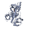
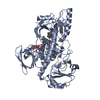
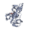
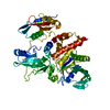
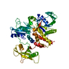
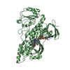
oxidoreductase activity, acting on paired donors, with incorporation or reduction of molecular oxygen, NAD(P)H as one donor, and incorporation of one atom of oxygen / FAD binding / methyltransferase activity / methylation Similarity search - Function
Mass: 18.015 Da / Num. of mol.: 353 / Source method: isolated from a natural source / Formula: H2O
-
Experimental details
-
Experiment
Experiment
Method: X-RAY DIFFRACTION / Number of used crystals: 1
-
Sample preparation
Crystal
Density Matthews: 2.59 Å3/Da / Density % sol: 52.48 %
Crystal grow
Temperature: 298 K / Method: vapor diffusion, hanging drop / pH: 7.4 Details: 1.5 microliters of RebC (9 mg/mL in 150 mM NaCl, 10% glycerol, 25 mM HEPES pH 7.5) was incubated with 0.35 microliters of guanidine-HCl for 30 seconds, followed by addition of 1.5 ...Details: 1.5 microliters of RebC (9 mg/mL in 150 mM NaCl, 10% glycerol, 25 mM HEPES pH 7.5) was incubated with 0.35 microliters of guanidine-HCl for 30 seconds, followed by addition of 1.5 microliters of precipitant solution (19% PEG-8000, 0.1 M HEPES pH 7.4), without mixing, at room temperature and sealed over a precipitant well solution. Immediately after set up, crystal trays were placed on a gel shaker and then, after 12 hours, transferred to a storage space in vibration-isolation. A crystal was then soaked in 19% PEG-8000, 0.1 M HEPES pH 7.4, and 5 mM chromopyrrolic acid for 1 week. The crystal was then soaked for 5 seconds in a cryogenic solution containing 19% PEG-8000, 0.1 M HEPES pH 7.4, 20% glycerol, and 5 mM chromopyrrolic acid and then flash-frozen in liquid nitrogen, VAPOR DIFFUSION, HANGING DROP, temperature 298K
In the structure databanks used in Yorodumi, some data are registered as the other names, "COVID-19 virus" and "2019-nCoV". Here are the details of the virus and the list of structure data.
Jan 31, 2019. EMDB accession codes are about to change! (news from PDBe EMDB page)
EMDB accession codes are about to change! (news from PDBe EMDB page)
The allocation of 4 digits for EMDB accession codes will soon come to an end. Whilst these codes will remain in use, new EMDB accession codes will include an additional digit and will expand incrementally as the available range of codes is exhausted. The current 4-digit format prefixed with “EMD-” (i.e. EMD-XXXX) will advance to a 5-digit format (i.e. EMD-XXXXX), and so on. It is currently estimated that the 4-digit codes will be depleted around Spring 2019, at which point the 5-digit format will come into force.
The EM Navigator/Yorodumi systems omit the EMD- prefix.
Related info.:Q: What is EMD? / ID/Accession-code notation in Yorodumi/EM Navigator
Yorodumi is a browser for structure data from EMDB, PDB, SASBDB, etc.
This page is also the successor to EM Navigator detail page, and also detail information page/front-end page for Omokage search.
The word "yorodu" (or yorozu) is an old Japanese word meaning "ten thousand". "mi" (miru) is to see.
Related info.:EMDB / PDB / SASBDB / Comparison of 3 databanks / Yorodumi Search / Aug 31, 2016. New EM Navigator & Yorodumi / Yorodumi Papers / Jmol/JSmol / Function and homology information / Changes in new EM Navigator and Yorodumi
 Movie
Movie Controller


 Open data
 Basic information
 Components
 Keywords
 Function and homology information
 Lechevalieria aerocolonigenes (bacteria)
 X-RAY DIFFRACTION /
  Authors
 Citation
 Structure visualization
 Downloads & links
 Other downloads


 PDBj
PDBj Assembly

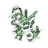
 Mass: 785.550 Da / Num. of mol.: 2 / Source method: obtained synthetically / Formula: C27H33N9O15P2 / Comment: FAD*YM
 Mass: 355.346 Da / Num. of mol.: 2 / Source method: obtained synthetically / Formula: C21H13N3O3
 Mass: 18.015 Da / Num. of mol.: 353 / Source method: isolated from a natural source / Formula: H2O
 Sample preparation
 / Beamline: 24-ID-C / Wavelength: 0.9795 Å
 Processing