 Movie
Movie Controller

[English] 日本語
 Yorodumi
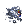
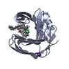
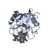
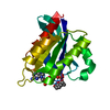
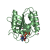
- PDB-2qz3: Crystal structure of a glycoside hydrolase family 11 xylanase fro... -
+ Open data

- Basic information
| Entry | Database: PDB / ID: 2qz3 | |||||||||
|---|---|---|---|---|---|---|---|---|---|---|
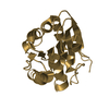
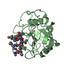
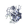
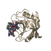
| Title | Crystal structure of a glycoside hydrolase family 11 xylanase from Bacillus subtilis in complex with xylotetraose | |||||||||
 Components Components | Endo-1,4-beta-xylanase A | |||||||||
 Keywords Keywords | HYDROLASE / glycoside hydrolase / xylanase / Glycosidase / Xylan degradation | |||||||||
| Function / homology |  Function and homology information Function and homology informationendo-1,4-beta-xylanase / endo-1,4-beta-xylanase activity / xylan catabolic process Similarity search - Function | |||||||||
| Biological species |  | |||||||||
| Method |  X-RAY DIFFRACTION / SYNCHROTRON / MOLECULAR REPLACEMENT / Resolution: 1.8 Å X-RAY DIFFRACTION / SYNCHROTRON / MOLECULAR REPLACEMENT / Resolution: 1.8 Å | |||||||||
 Authors Authors | Vandermarliere, E. / Bourgois, T.M. / Strelkov, S.V. / Delcour, J.A. / Courtin, C.M. / Rabijns, A. | |||||||||
 Citation Citation | Journal: Biochem.J. / Year: 2008 Title: Crystallographic analysis shows substrate binding at the -3 to +1 active-site subsites and at the surface of glycoside hydrolase family 11 endo-1,4-beta-xylanases. Authors: Vandermarliere, E. / Bourgois, T.M. / Rombouts, S. / Van Campenhout, S. / Volckaert, G. / Strelkov, S.V. / Delcour, J.A. / Rabijns, A. / Courtin, C.M. | |||||||||
| History |
|
- Structure visualization
| Structure viewer | Molecule: MolmilJmol/JSmol |
|---|
- Downloads & links
-Download
| PDBx/mmCIF format | 2qz3.cif.gz | 94.1 KB | Display | PDBx/mmCIF format |
|---|---|---|---|---|
| PDB format | pdb2qz3.ent.gz | 71.1 KB | Display | PDB format |
| PDBx/mmJSON format | 2qz3.json.gz | Tree view | PDBx/mmJSON format | |
| Others |  Other downloads Other downloads |
-Validation report
| Arichive directory | https://data.pdbj.org/pub/pdb/validation_reports/qz/2qz3ftp://data.pdbj.org/pub/pdb/validation_reports/qz/2qz3 | HTTPS FTP |
|---|
-Related structure data
| Related structure data |  2qz2C  2z79C  1bcxS S: Starting model for refinement C: citing same article ( |
|---|---|
| Similar structure data |

-Links
 PDBj
PDBj


- Assembly
| Deposited unit | 
| ||||||||
|---|---|---|---|---|---|---|---|---|---|
| 1 | 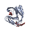
| ||||||||
| 2 | 
| ||||||||
| Unit cell |
|
-Components
| #1: Protein | Mass: 20336.943 Da / Num. of mol.: 2 / Mutation: E172A Source method: isolated from a genetically manipulated source Source: (gene. exp.) #2: Polysaccharide | beta-D-xylopyranose-(1-4)-beta-D-xylopyranose-(1-4)-beta-D-xylopyranose / 4beta-beta-xylotriose   Source method: isolated from a genetically manipulated source Details: oligosaccharide / References: 4beta-beta-xylotriose #3: Chemical | ChemComp-ACY / |   Mass: 60.052 Da / Num. of mol.: 1 / Source method: obtained synthetically / Formula: C2H4O2 Mass: 60.052 Da / Num. of mol.: 1 / Source method: obtained synthetically / Formula: C2H4O2#4: Water | ChemComp-HOH / |  Mass: 18.015 Da / Num. of mol.: 295 / Source method: isolated from a natural source / Formula: H2O Mass: 18.015 Da / Num. of mol.: 295 / Source method: isolated from a natural source / Formula: H2O |
|---|
-Experimental details
-Experiment
| Experiment | Method: X-RAY DIFFRACTION / Number of used crystals: 1 |
|---|
- Sample preparation
| Crystal | Density Matthews: 2.1 Å3/Da / Density % sol: 41.52 % |
|---|---|
| Crystal grow | Temperature: 277 K / Method: vapor diffusion, hanging drop / pH: 8.5 Details: 0.2M ammonium sulphate, 0.1M Tris-HCl pH 8.5, 30% isopropanol, VAPOR DIFFUSION, HANGING DROP, temperature 277K |
-Data collection
| Diffraction | Mean temperature: 100 K |
|---|---|
| Diffraction source | Source: SYNCHROTRON / Site: EMBL/DESY, HAMBURG  / Beamline: BW7A / Wavelength: 0.9762 Å / Beamline: BW7A / Wavelength: 0.9762 Å |
| Detector | Type: MAR CCD 165 mm / Detector: CCD / Date: Mar 12, 2006 |
| Radiation | Monochromator: Fixed exit double crystal Si [111], horizontally focussing Protocol: SINGLE WAVELENGTH / Monochromatic (M) / Laue (L): M / Scattering type: x-ray |
| Radiation wavelength | Wavelength: 0.9762 Å / Relative weight: 1 |
| Reflection | Resolution: 1.8→20 Å / Num. all: 31423 / Num. obs: 30942 / % possible obs: 99.4 % / Observed criterion σ(F): 2 / Observed criterion σ(I): 1.4 / Rsym value: 0.044 / Net I/σ(I): 23.3 |
| Reflection shell | Resolution: 1.8→1.83 Å / Mean I/σ(I) obs: 11.5 / Num. unique all: 1547 / Rsym value: 0.089 / % possible all: 99.9 |
- Processing
| Software |
| ||||||||||||||||||||||||||||||||||||||||||||||||||||||||||||||||||||||||||||||||||||||||||
|---|---|---|---|---|---|---|---|---|---|---|---|---|---|---|---|---|---|---|---|---|---|---|---|---|---|---|---|---|---|---|---|---|---|---|---|---|---|---|---|---|---|---|---|---|---|---|---|---|---|---|---|---|---|---|---|---|---|---|---|---|---|---|---|---|---|---|---|---|---|---|---|---|---|---|---|---|---|---|---|---|---|---|---|---|---|---|---|---|---|---|---|
| Refinement | Method to determine structure: MOLECULAR REPLACEMENT Starting model: 1BCX Resolution: 1.8→19.91 Å / Cor.coef. Fo:Fc: 0.963 / Cor.coef. Fo:Fc free: 0.94 / SU B: 2.029 / SU ML: 0.066 / Cross valid method: THROUGHOUT / σ(F): 0 / ESU R: 0.131 / ESU R Free: 0.122 / Stereochemistry target values: MAXIMUM LIKELIHOOD / Details: HYDROGENS HAVE BEEN ADDED IN THE RIDING POSITIONS
| ||||||||||||||||||||||||||||||||||||||||||||||||||||||||||||||||||||||||||||||||||||||||||
| Solvent computation | Ion probe radii: 0.8 Å / Shrinkage radii: 0.8 Å / VDW probe radii: 1.4 Å / Solvent model: MASK | ||||||||||||||||||||||||||||||||||||||||||||||||||||||||||||||||||||||||||||||||||||||||||
| Displacement parameters | Biso mean: 14.35 Å2
| ||||||||||||||||||||||||||||||||||||||||||||||||||||||||||||||||||||||||||||||||||||||||||
| Refinement step | Cycle: LAST / Resolution: 1.8→19.91 Å
| ||||||||||||||||||||||||||||||||||||||||||||||||||||||||||||||||||||||||||||||||||||||||||
| Refine LS restraints |
| ||||||||||||||||||||||||||||||||||||||||||||||||||||||||||||||||||||||||||||||||||||||||||
| LS refinement shell | Resolution: 1.802→1.849 Å / Total num. of bins used: 20
|
