BIOMOLECULE: 1, 2, 3 SEE REMARK 350 FOR THE AUTHOR PROVIDED AND PROGRAM GENERATED ASSEMBLY ... BIOMOLECULE: 1, 2, 3 SEE REMARK 350 FOR THE AUTHOR PROVIDED AND PROGRAM GENERATED ASSEMBLY INFORMATION FOR THE STRUCTURE IN THIS ENTRY. THIS ENTRY CONTAINS THE CRYSTALLOGRAPHIC ASYMMETRIC UNIT WHICH CONSISTS OF 3 CHAINS BUT THEY DO NOT FORM OLIGOMERS BASED ON CRYSTAL PACKING ANALYSIS. SIZE EXCLUSION CHROMATOGRAPHY SUPPORTS THE ASSIGNMENT OF A MONOMER AS THE SIGNIFICANT OLIGOMERIZATION STATE.
Remark 999
SEQUENCE THE CONSTRUCT WAS EXPRESSED WITH A PURIFICATION TAG MGSDKIHHHHHHENLYFQG. THE TAG WAS ... SEQUENCE THE CONSTRUCT WAS EXPRESSED WITH A PURIFICATION TAG MGSDKIHHHHHHENLYFQG. THE TAG WAS REMOVED WITH TEV PROTEASE LEAVING ONLY A GLYCINE FOLLOWED BY THE TARGET SEQUENCE.
Component-ID: 1 / Ens-ID: 1 / Beg auth comp-ID: GLN / Beg label comp-ID: GLN / End auth comp-ID: PRO / End label comp-ID: PRO / Refine code: 6 / Auth seq-ID: 7 - 271 / Label seq-ID: 8 - 272
Dom-ID
Auth asym-ID
Label asym-ID
1
A
A
2
B
B
3
C
C
Details
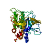
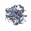
THIS ENTRY CONTAINS THE CRYSTALLOGRAPHIC ASYMMETRIC UNIT WHICH CONSISTS OF 3 CHAINS BUT THEY DO NOT FORM OLIGOMERS BASED ON CRYSTAL PACKING ANALYSIS. SIZE EXCLUSION CHROMATOGRAPHY SUPPORTS THE ASSIGNMENT OF A MONOMER AS THE SIGNIFICANT OLIGOMERIZATION STATE.
-
Components
#1: Protein
Uncharacterizedprotein
Mass: 30339.539 Da / Num. of mol.: 3 Source method: isolated from a genetically manipulated source Source: (gene. exp.) Shewanella amazonensis (bacteria) / Strain: SB2B / Gene: YP_926603.1, Sama_0725 / Plasmid: speedET / Production host: Escherichia coli (E. coli) / Strain (production host): HK100 / References: UniProt: A1S3H7
Type: MARMOSAIC 325 mm CCD / Detector: CCD / Date: Jun 22, 2007 / Details: Flat mirror (vertical focusing)
Radiation
Monochromator: Single crystal Si(111) bent (horizontal focusing) Protocol: MAD / Monochromatic (M) / Laue (L): M / Scattering type: x-ray
Radiation wavelength
ID
Wavelength (Å)
Relative weight
1
0.91837
1
2
0.97944
1
Reflection
Resolution: 2→29.63 Å / Num. obs: 57061 / % possible obs: 92.8 % / Redundancy: 3.36 % / Biso Wilson estimate: 20.14 Å2 / Rmerge(I) obs: 0.107 / Net I/σ(I): 7.41
Reflection shell
Resolution (Å)
Rmerge(I) obs
Mean I/σ(I) obs
Num. measured obs
Diffraction-ID
% possible all
2-2.07
0.518
1.87
19189
1
99.5
2.07-2.15
0.434
2.3
19546
1
99.8
2.15-2.25
0.368
2.7
10720
1
53.8
2.25-2.37
0.303
3.3
18632
1
91.7
2.37-2.52
0.252
4.1
20909
1
99.9
2.52-2.71
0.198
5.2
20392
1
99.9
2.71-2.99
0.133
7.2
21634
1
99.8
2.99-3.42
0.078
11.1
20982
1
99.9
3.42-4.3
0.05
15.5
17921
1
85.7
4.3-29.63
0.035
19.4
21955
1
99.6
-
Phasing
Phasing
Method: MAD
-
Processing
Software
Name
Version
Classification
NB
REFMAC
5.2.0019
refinement
PHENIX
refinement
SHELX
phasing
MolProbity
3beta29
modelbuilding
XSCALE
datascaling
PDB_EXTRACT
2
dataextraction
MAR345
CCD
datacollection
XDS
datareduction
SHELXD
phasing
SHARP
phasing
Refinement
Method to determine structure: MAD / Resolution: 2→29.63 Å / Cor.coef. Fo:Fc: 0.959 / Cor.coef. Fo:Fc free: 0.929 / SU B: 7.307 / SU ML: 0.108 / TLS residual ADP flag: LIKELY RESIDUAL / Cross valid method: THROUGHOUT / σ(F): 0 / ESU R: 0.172 / ESU R Free: 0.159 Stereochemistry target values: MAXIMUM LIKELIHOOD WITH PHASES Details: 1. HYDROGENS HAVE BEEN ADDED IN THE RIDING POSITIONS. 2. A MET-INHIBITION PROTOCOL WAS USED FOR SELENOMETHIONINE INCORPORATION DURING PROTEIN EXPRESSION. THE OCCUPANCY OF THE SE ATOMS IN THE ...Details: 1. HYDROGENS HAVE BEEN ADDED IN THE RIDING POSITIONS. 2. A MET-INHIBITION PROTOCOL WAS USED FOR SELENOMETHIONINE INCORPORATION DURING PROTEIN EXPRESSION. THE OCCUPANCY OF THE SE ATOMS IN THE MSE RESIDUES WAS REDUCED TO 0.75 TO ACCOUNT FOR THE REDUCED SCATTERING POWER DUE TO PARTIAL S-MET INCORPORATION. 3. ATOM RECORD CONTAINS RESIDUAL B FACTORS ONLY. 4. RESIDUES A1-A6, A273-A274, B1-B5, B273-B274, AND C1-C5 ARE DISORDERED AND HAVE NOT BEEN MODELLED. 4. TEN SULFATE ANIONS AND TWELVE GLYCEROL MOLECULES HAVE BEEN MODELED IN THE SOLVENT STRUCTURE. 5. DIFFRACTION DATA IMAGES WERE PROCESSED BY MASKING OUT REFLECTIONS IN RESOLUTION RANGES 3.89A-3.87A, 3.70A-3.64A, 2.26A-2.24A AND 2.24A-2.21A CORRESPONDING TO ICE RINGS.
Rfactor
Num. reflection
% reflection
Selection details
Rfree
0.213
2870
5 %
RANDOM
Rwork
0.161
-
-
-
all
0.164
-
-
-
obs
0.164
57042
93.32 %
-
Solvent computation
Ion probe radii: 0.8 Å / Shrinkage radii: 0.8 Å / VDW probe radii: 1.2 Å / Solvent model: MASK
In the structure databanks used in Yorodumi, some data are registered as the other names, "COVID-19 virus" and "2019-nCoV". Here are the details of the virus and the list of structure data.
Jan 31, 2019. EMDB accession codes are about to change! (news from PDBe EMDB page)
EMDB accession codes are about to change! (news from PDBe EMDB page)
The allocation of 4 digits for EMDB accession codes will soon come to an end. Whilst these codes will remain in use, new EMDB accession codes will include an additional digit and will expand incrementally as the available range of codes is exhausted. The current 4-digit format prefixed with “EMD-” (i.e. EMD-XXXX) will advance to a 5-digit format (i.e. EMD-XXXXX), and so on. It is currently estimated that the 4-digit codes will be depleted around Spring 2019, at which point the 5-digit format will come into force.
The EM Navigator/Yorodumi systems omit the EMD- prefix.
Related info.:Q: What is EMD? / ID/Accession-code notation in Yorodumi/EM Navigator
Yorodumi is a browser for structure data from EMDB, PDB, SASBDB, etc.
This page is also the successor to EM Navigator detail page, and also detail information page/front-end page for Omokage search.
The word "yorodu" (or yorozu) is an old Japanese word meaning "ten thousand". "mi" (miru) is to see.
Related info.:EMDB / PDB / SASBDB / Comparison of 3 databanks / Yorodumi Search / Aug 31, 2016. New EM Navigator & Yorodumi / Yorodumi Papers / Jmol/JSmol / Function and homology information / Changes in new EM Navigator and Yorodumi
 Movie
Movie Controller

 Yorodumi
 Open data
 Basic information
 Components
 Keywords
 Function and homology information
 Shewanella amazonensis (bacteria)
 X-RAY DIFFRACTION /
  Authors
 Citation
 Structure visualization
 Downloads & links
 Other downloads


 PDBj
PDBj
 Assembly
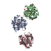


 Mass: 96.063 Da / Num. of mol.: 10 / Source method: obtained synthetically / Formula: SO4
 Mass: 92.094 Da / Num. of mol.: 12 / Source method: obtained synthetically / Formula: C3H8O3
 Mass: 18.015 Da / Num. of mol.: 526 / Source method: isolated from a natural source / Formula: H2O
 Sample preparation
 / Beamline: BL11-1 / Wavelength: 0.91837, 0.97944
 Processing