 Movie
Movie Controller

[English] 日本語
 Yorodumi
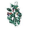
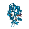
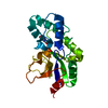
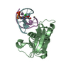
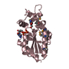
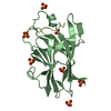
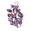
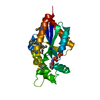
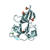
- PDB-2qt0: Human nicotinamide riboside kinase 1 in complex with nicotinamide... -
+ Open data

- Basic information
| Entry | Database: PDB / ID: 2qt0 | ||||||
|---|---|---|---|---|---|---|---|
| Title | Human nicotinamide riboside kinase 1 in complex with nicotinamide riboside and an ATP analogue | ||||||
 Components Components | Nicotinamide riboside kinase 1 | ||||||
 Keywords Keywords | TRANSFERASE / non-protein kinase / NAD+ / nicotinamide riboside / nrk1 / nicotinamide riboside kinase activity / nicotinic acid riboside kinase activity / NAD biosynthesis / pyridine nucleotide biosynthesis / ATP analogue / STRUCTURAL GENOMICS / STRUCTURAL GENOMICS CONSORTIUM / SGC / Alternative splicing / ATP-binding / Nucleotide-binding | ||||||
| Function / homology |  Function and homology information Function and homology informationnicotinate riboside kinase / nicotinamide riboside metabolic process / nicotinate riboside kinase activity / ribosylnicotinamide kinase / ribosylnicotinamide kinase activity / NAD+ biosynthetic process via the salvage pathway / nicotinate metabolic process / NADP+ biosynthetic process / Nicotinate metabolism / ATP binding ...nicotinate riboside kinase / nicotinamide riboside metabolic process / nicotinate riboside kinase activity / ribosylnicotinamide kinase / ribosylnicotinamide kinase activity / NAD+ biosynthetic process via the salvage pathway / nicotinate metabolic process / NADP+ biosynthetic process / Nicotinate metabolism / ATP binding / metal ion binding / cytoplasm / cytosol Similarity search - Function | ||||||
| Biological species |  Homo sapiens (human) Homo sapiens (human) | ||||||
| Method |  X-RAY DIFFRACTION / MOLECULAR REPLACEMENT / molecular replacement / Resolution: 1.92 Å X-RAY DIFFRACTION / MOLECULAR REPLACEMENT / molecular replacement / Resolution: 1.92 Å | ||||||
 Authors Authors | Rabeh, W.M. / Tempel, W. / Nedyalkova, L. / Landry, R. / Arrowsmith, C.H. / Edwards, A.M. / Sundstrom, M. / Weigelt, J. / Bochkarev, A. / Brenner, C. ...Rabeh, W.M. / Tempel, W. / Nedyalkova, L. / Landry, R. / Arrowsmith, C.H. / Edwards, A.M. / Sundstrom, M. / Weigelt, J. / Bochkarev, A. / Brenner, C. / Park, H. / Structural Genomics Consortium (SGC) | ||||||
 Citation Citation | Journal: Plos Biol. / Year: 2007 Title: Nicotinamide Riboside Kinase Structures Reveal New Pathways to NAD(+). Authors: Tempel, W. / Rabeh, W.M. / Bogan, K.L. / Belenky, P. / Wojcik, M. / Seidle, H.F. / Nedyalkova, L. / Yang, T. / Sauve, A.A. / Park, H.W. / Brenner, C. | ||||||
| History |
| ||||||
| Remark 300 | BIOMOLECULE: 1 SEE REMARK 350 FOR THE PROGRAM GENERATED ASSEMBLY INFORMATION FOR THE STRUCTURE IN ... BIOMOLECULE: 1 SEE REMARK 350 FOR THE PROGRAM GENERATED ASSEMBLY INFORMATION FOR THE STRUCTURE IN THIS ENTRY. AUTHORS STATE THAT THE BIOLOGICAL UNIT OF THIS POLYPEPTIDE IS UNKNOWN. |
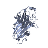
- Structure visualization
| Structure viewer | Molecule: MolmilJmol/JSmol |
|---|
- Downloads & links
-Download
| PDBx/mmCIF format | 2qt0.cif.gz | 56.9 KB | Display | PDBx/mmCIF format |
|---|---|---|---|---|
| PDB format | pdb2qt0.ent.gz | 38.1 KB | Display | PDB format |
| PDBx/mmJSON format | 2qt0.json.gz | Tree view | PDBx/mmJSON format | |
| Others |  Other downloads Other downloads |
-Validation report
| Arichive directory | https://data.pdbj.org/pub/pdb/validation_reports/qt/2qt0ftp://data.pdbj.org/pub/pdb/validation_reports/qt/2qt0 | HTTPS FTP |
|---|
-Related structure data
| Related structure data | 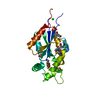 2p0eSC 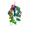 2qsyC 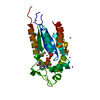 2qszC 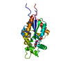 2qt1C C: citing same article ( S: Starting model for refinement |
|---|---|
| Similar structure data |
-Links
 PDBj
PDBj


- Assembly
| Deposited unit | 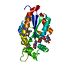
| ||||||||
|---|---|---|---|---|---|---|---|---|---|
| 1 |
| ||||||||
| Unit cell |
| ||||||||
| Details | not known |
-Components
| #1: Protein | Mass: 24373.371 Da / Num. of mol.: 1 Source method: isolated from a genetically manipulated source Source: (gene. exp.) Homo sapiens (human) / Gene: NRK1, C9orf95 / Plasmid: p28a-LIC / Production host:  References: UniProt: Q9NWW6, Transferases; Transferring phosphorus-containing groups; Phosphotransferases with an alcohol group as acceptor |
|---|---|
| #2: Chemical | ChemComp-MG /   Mass: 24.305 Da / Num. of mol.: 1 / Source method: obtained synthetically / Formula: Mg Mass: 24.305 Da / Num. of mol.: 1 / Source method: obtained synthetically / Formula: Mg |
| #3: Chemical | ChemComp-ANP /   Mass: 506.196 Da / Num. of mol.: 1 / Source method: obtained synthetically / Formula: C10H17N6O12P3 / Comment: AMP-PNP, energy-carrying molecule analogue*YM Mass: 506.196 Da / Num. of mol.: 1 / Source method: obtained synthetically / Formula: C10H17N6O12P3 / Comment: AMP-PNP, energy-carrying molecule analogue*YM |
| #4: Chemical | ChemComp-NNR / 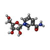  Mass: 255.247 Da / Num. of mol.: 1 / Source method: obtained synthetically / Formula: C11H15N2O5 Mass: 255.247 Da / Num. of mol.: 1 / Source method: obtained synthetically / Formula: C11H15N2O5 |
| #5: Water | ChemComp-HOH /  Mass: 18.015 Da / Num. of mol.: 35 / Source method: isolated from a natural source / Formula: H2O Mass: 18.015 Da / Num. of mol.: 35 / Source method: isolated from a natural source / Formula: H2O |
| Has protein modification | Y |
-Experimental details
-Experiment
| Experiment | Method: X-RAY DIFFRACTION / Number of used crystals: 1 |
|---|
- Sample preparation
| Crystal | Density Matthews: 2.16 Å3/Da / Density % sol: 43.13 % |
|---|---|
| Crystal grow | Temperature: 291 K / Method: vapor diffusion / pH: 8 Details: 35% PEG 2000 MME, 0.1M Tris-HCl. The protein solution (40mg/mL) contained 0.01M Nicotinamide riboside, 0.01M AMPPNP and 0.02M Magnesium chloride, pH 8.0, VAPOR DIFFUSION, temperature 291K |
-Data collection
| Diffraction | Mean temperature: 100 K | ||||||||||||||||||||||||||||||||||||||||||||||||||||||||||||||||||
|---|---|---|---|---|---|---|---|---|---|---|---|---|---|---|---|---|---|---|---|---|---|---|---|---|---|---|---|---|---|---|---|---|---|---|---|---|---|---|---|---|---|---|---|---|---|---|---|---|---|---|---|---|---|---|---|---|---|---|---|---|---|---|---|---|---|---|---|
| Diffraction source | Source: ROTATING ANODE / Type: RIGAKU FR-E+ SUPERBRIGHT / Wavelength: 1.5418 Å | ||||||||||||||||||||||||||||||||||||||||||||||||||||||||||||||||||
| Detector | Type: RIGAKU RAXIS / Detector: IMAGE PLATE / Date: Mar 1, 2007 | ||||||||||||||||||||||||||||||||||||||||||||||||||||||||||||||||||
| Radiation | Protocol: SINGLE WAVELENGTH / Monochromatic (M) / Laue (L): M / Scattering type: x-ray | ||||||||||||||||||||||||||||||||||||||||||||||||||||||||||||||||||
| Radiation wavelength | Wavelength: 1.5418 Å / Relative weight: 1 | ||||||||||||||||||||||||||||||||||||||||||||||||||||||||||||||||||
| Reflection | Resolution: 1.92→30 Å / Num. obs: 31229 / % possible obs: 100 % / Redundancy: 6.5 % / Rmerge(I) obs: 0.086 / Χ2: 1.911 / Net I/σ(I): 10.7 | ||||||||||||||||||||||||||||||||||||||||||||||||||||||||||||||||||
| Reflection shell |
|
-Phasing
| Phasing | Method: molecular replacement | |||||||||
|---|---|---|---|---|---|---|---|---|---|---|
| Phasing MR |
|
- Processing
| Software |
| |||||||||||||||||||||||||||||||||||||||||||||||||||||||||||||||||||||||||||||||||||||||||||||||||||||||||||||||||||||||||||||||||||||||||||||||||||
|---|---|---|---|---|---|---|---|---|---|---|---|---|---|---|---|---|---|---|---|---|---|---|---|---|---|---|---|---|---|---|---|---|---|---|---|---|---|---|---|---|---|---|---|---|---|---|---|---|---|---|---|---|---|---|---|---|---|---|---|---|---|---|---|---|---|---|---|---|---|---|---|---|---|---|---|---|---|---|---|---|---|---|---|---|---|---|---|---|---|---|---|---|---|---|---|---|---|---|---|---|---|---|---|---|---|---|---|---|---|---|---|---|---|---|---|---|---|---|---|---|---|---|---|---|---|---|---|---|---|---|---|---|---|---|---|---|---|---|---|---|---|---|---|---|---|---|---|---|
| Refinement | Method to determine structure: MOLECULAR REPLACEMENT Starting model: PDB entry 2P0E Resolution: 1.92→30 Å / Cor.coef. Fo:Fc: 0.953 / Cor.coef. Fo:Fc free: 0.94 / WRfactor Rfree: 0.226 / WRfactor Rwork: 0.191 / Cross valid method: THROUGHOUT / σ(F): 0 / ESU R: 0.162 / ESU R Free: 0.147 / Stereochemistry target values: MAXIMUM LIKELIHOOD Details: 1. The Bijvoet differences were used for phasing. 2. Arp/warp, coot, prodrg, molprobity programs have also been used in refinement. 3. HYDROGENS HAVE BEEN ADDED IN THE RIDING POSITIONS.
| |||||||||||||||||||||||||||||||||||||||||||||||||||||||||||||||||||||||||||||||||||||||||||||||||||||||||||||||||||||||||||||||||||||||||||||||||||
| Solvent computation | Ion probe radii: 0.8 Å / Shrinkage radii: 0.8 Å / VDW probe radii: 1.4 Å / Solvent model: MASK | |||||||||||||||||||||||||||||||||||||||||||||||||||||||||||||||||||||||||||||||||||||||||||||||||||||||||||||||||||||||||||||||||||||||||||||||||||
| Displacement parameters | Biso mean: 27.811 Å2
| |||||||||||||||||||||||||||||||||||||||||||||||||||||||||||||||||||||||||||||||||||||||||||||||||||||||||||||||||||||||||||||||||||||||||||||||||||
| Refinement step | Cycle: LAST / Resolution: 1.92→30 Å
| |||||||||||||||||||||||||||||||||||||||||||||||||||||||||||||||||||||||||||||||||||||||||||||||||||||||||||||||||||||||||||||||||||||||||||||||||||
| Refine LS restraints |
| |||||||||||||||||||||||||||||||||||||||||||||||||||||||||||||||||||||||||||||||||||||||||||||||||||||||||||||||||||||||||||||||||||||||||||||||||||
| LS refinement shell | Refine-ID: X-RAY DIFFRACTION / Total num. of bins used: 20
|
