- PDB-2qrz: Cdc42 bound to GMP-PCP: Induced Fit by Effector is Required -
+
Open data
ID or keywords:
Loading...
-
Basic information
Entry
Database: PDB / ID: 2qrz
Title
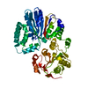
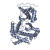
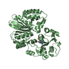
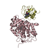
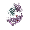
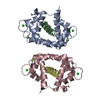
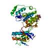
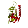
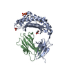
Cdc42 bound to GMP-PCP: Induced Fit by Effector is Required
Components
Cell division control protein 42 homolog precursor
Keywords
CELL CYCLE / G-domain fold / G protein / GTPase / Alternative splicing / GTP-binding / Lipoprotein / Membrane / Methylation / Nucleotide-binding / Prenylation
Function / homology
Function and homology information
GBD domain binding / positive regulation of pinocytosis / COG complex / endothelin receptor signaling pathway involved in heart process / cardiac neural crest cell migration involved in outflow tract morphogenesis / storage vacuole / dendritic cell migration / neuron fate determination / apolipoprotein A-I receptor binding / positive regulation of epithelial cell proliferation involved in lung morphogenesis ...GBD domain binding / positive regulation of pinocytosis / COG complex / endothelin receptor signaling pathway involved in heart process / cardiac neural crest cell migration involved in outflow tract morphogenesis / storage vacuole / dendritic cell migration / neuron fate determination / apolipoprotein A-I receptor binding / positive regulation of epithelial cell proliferation involved in lung morphogenesis / regulation of attachment of spindle microtubules to kinetochore / organelle transport along microtubule / positive regulation of pseudopodium assembly / Inactivation of CDC42 and RAC1 / cardiac conduction system development / host-mediated perturbation of viral process / leading edge membrane / regulation of filopodium assembly / neuropilin signaling pathway / establishment of Golgi localization / filopodium assembly / cell junction assembly / dendritic spine morphogenesis / establishment of epithelial cell apical/basal polarity / adherens junction organization / GTP-dependent protein binding / thioesterase binding / regulation of lamellipodium assembly / regulation of stress fiber assembly / embryonic heart tube development / RHO GTPases activate KTN1 / DCC mediated attractive signaling / CD28 dependent Vav1 pathway / regulation of postsynapse organization / positive regulation of filopodium assembly / Wnt signaling pathway, planar cell polarity pathway / phagocytosis, engulfment / RHOV GTPase cycle / Myogenesis / nuclear migration / regulation of mitotic nuclear division / small GTPase-mediated signal transduction / positive regulation of cytokinesis / spindle midzone / RHOJ GTPase cycle / heart contraction / RHOQ GTPase cycle / establishment of cell polarity / Golgi organization / RHOU GTPase cycle / establishment or maintenance of cell polarity / RHO GTPases activate PAKs / CDC42 GTPase cycle / macrophage differentiation / RHOG GTPase cycle / RAC3 GTPase cycle / RAC2 GTPase cycle / RHO GTPases Activate WASPs and WAVEs / negative regulation of protein-containing complex assembly / RHO GTPases activate IQGAPs / GPVI-mediated activation cascade / positive regulation of lamellipodium assembly / phagocytic vesicle / positive regulation of stress fiber assembly / RAC1 GTPase cycle / EPHB-mediated forward signaling / positive regulation of substrate adhesion-dependent cell spreading / substantia nigra development / Gene and protein expression by JAK-STAT signaling after Interleukin-12 stimulation / actin filament organization / integrin-mediated signaling pathway / small monomeric GTPase / regulation of actin cytoskeleton organization / FCGR3A-mediated phagocytosis / filopodium / EGFR downregulation / RHO GTPases Activate Formins / Regulation of actin dynamics for phagocytic cup formation / MAPK6/MAPK4 signaling / VEGFA-VEGFR2 Pathway / cellular response to type II interferon / endocytosis / apical part of cell / cytoplasmic ribonucleoprotein granule / cell-cell junction / mitotic spindle / G beta:gamma signalling through CDC42 / intracellular protein localization / ubiquitin protein ligase activity / Factors involved in megakaryocyte development and platelet production / positive regulation of cell growth / actin cytoskeleton organization / G protein activity / midbody / positive regulation of MAPK cascade / positive regulation of phosphatidylinositol 3-kinase/protein kinase B signal transduction / neuron projection / postsynapse / positive regulation of cell migration / Golgi membrane Similarity search - Function
Cdc42 / Small GTPase Rho / Small GTPase Rho domain profile. / Rho (Ras homology) subfamily of Ras-like small GTPases / Ras subfamily of RAS small GTPases / Small GTPase / Ras family / Rab subfamily of small GTPases / Small GTP-binding protein domain / P-loop containing nucleotide triphosphate hydrolases ...Cdc42 / Small GTPase Rho / Small GTPase Rho domain profile. / Rho (Ras homology) subfamily of Ras-like small GTPases / Ras subfamily of RAS small GTPases / Small GTPase / Ras family / Rab subfamily of small GTPases / Small GTP-binding protein domain / P-loop containing nucleotide triphosphate hydrolases / Rossmann fold / P-loop containing nucleoside triphosphate hydrolase / 3-Layer(aba) Sandwich / Alpha Beta Similarity search - Domain/homology
PHOSPHOMETHYLPHOSPHONIC ACID GUANYLATE ESTER / Cell division control protein 42 homolog Similarity search - Component
Mass: 18.015 Da / Num. of mol.: 115 / Source method: isolated from a natural source / Formula: H2O
Has protein modification
Y
-
Experimental details
-
Experiment
Experiment
Method: X-RAY DIFFRACTION / Number of used crystals: 1
-
Sample preparation
Crystal
Density Matthews: 2.95 Å3/Da / Density % sol: 58.29 %
Crystal grow
Temperature: 277 K / Method: vapor diffusion, hanging drop / pH: 6 Details: 12% PEG 6K, 100 mM Na Acetate, 50 mM MES, pH 6.0, 100 mM Ammonium Sulfate, VAPOR DIFFUSION, HANGING DROP, temperature 277K
In the structure databanks used in Yorodumi, some data are registered as the other names, "COVID-19 virus" and "2019-nCoV". Here are the details of the virus and the list of structure data.
Jan 31, 2019. EMDB accession codes are about to change! (news from PDBe EMDB page)
EMDB accession codes are about to change! (news from PDBe EMDB page)
The allocation of 4 digits for EMDB accession codes will soon come to an end. Whilst these codes will remain in use, new EMDB accession codes will include an additional digit and will expand incrementally as the available range of codes is exhausted. The current 4-digit format prefixed with “EMD-” (i.e. EMD-XXXX) will advance to a 5-digit format (i.e. EMD-XXXXX), and so on. It is currently estimated that the 4-digit codes will be depleted around Spring 2019, at which point the 5-digit format will come into force.
The EM Navigator/Yorodumi systems omit the EMD- prefix.
Related info.:Q: What is EMD? / ID/Accession-code notation in Yorodumi/EM Navigator
Yorodumi is a browser for structure data from EMDB, PDB, SASBDB, etc.
This page is also the successor to EM Navigator detail page, and also detail information page/front-end page for Omokage search.
The word "yorodu" (or yorozu) is an old Japanese word meaning "ten thousand". "mi" (miru) is to see.
Related info.:EMDB / PDB / SASBDB / Comparison of 3 databanks / Yorodumi Search / Aug 31, 2016. New EM Navigator & Yorodumi / Yorodumi Papers / Jmol/JSmol / Function and homology information / Changes in new EM Navigator and Yorodumi
 Movie
Movie Controller


 Open data
 Basic information
 Components
 Keywords
 Function and homology information
 Homo sapiens (human)
 X-RAY DIFFRACTION /
  Authors
 Citation
 Structure visualization
 Downloads & links
 Other downloads
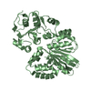
 PDBj
PDBj


 Assembly
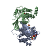


 Mass: 24.305 Da / Num. of mol.: 2 / Source method: obtained synthetically / Formula: Mg
 Mass: 96.063 Da / Num. of mol.: 1 / Source method: obtained synthetically / Formula: SO4
 Mass: 521.208 Da / Num. of mol.: 2 / Source method: obtained synthetically / Formula: C11H18N5O13P3 / Comment: GMP-PCP, energy-carrying molecule analogue*YM
 Mass: 18.015 Da / Num. of mol.: 115 / Source method: isolated from a natural source / Formula: H2O
 Sample preparation
 / Beamline: A1 / Wavelength: 0.978 Å
 Processing