 Movie
Movie Controller


+ Open data

- Basic information
| Entry | Database: PDB / ID: 2qoh | ||||||
|---|---|---|---|---|---|---|---|
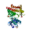
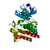
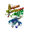
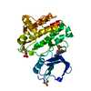
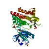
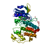
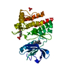
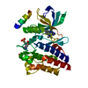
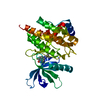
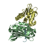
| Title | Crystal Structure of Abl kinase bound with PPY-A | ||||||
 Components Components | Proto-oncogene tyrosine-protein kinase ABL1 | ||||||
 Keywords Keywords | TRANSFERASE / Abl / kinase / inhibitor | ||||||
| Function / homology |  Function and homology information Function and homology informationRole of ABL in ROBO-SLIT signaling / HDR through Single Strand Annealing (SSA) / RHO GTPases Activate WASPs and WAVEs / MLL4 and MLL3 complexes regulate expression of PPARG target genes in adipogenesis and hepatic steatosis / Cyclin D associated events in G1 / Turbulent (oscillatory, disturbed) flow shear stress activates signaling by PIEZO1 and integrins in endothelial cells / Recruitment and ATM-mediated phosphorylation of repair and signaling proteins at DNA double strand breaks / protein localization to cytoplasmic microtubule plus-end / DNA conformation change / DN4 thymocyte differentiation ...Role of ABL in ROBO-SLIT signaling / HDR through Single Strand Annealing (SSA) / RHO GTPases Activate WASPs and WAVEs / MLL4 and MLL3 complexes regulate expression of PPARG target genes in adipogenesis and hepatic steatosis / Cyclin D associated events in G1 / Turbulent (oscillatory, disturbed) flow shear stress activates signaling by PIEZO1 and integrins in endothelial cells / Recruitment and ATM-mediated phosphorylation of repair and signaling proteins at DNA double strand breaks / protein localization to cytoplasmic microtubule plus-end / DNA conformation change / DN4 thymocyte differentiation / response to epinephrine / phospholipase C-inhibiting G protein-coupled receptor signaling pathway / podocyte apoptotic process / transitional one stage B cell differentiation / regulation of postsynaptic specialization assembly / positive regulation of phospholipase C/protein kinase C signal transduction / regulation of modification of synaptic structure / delta-catenin binding / cerebellum morphogenesis / RUNX1 regulates transcription of genes involved in differentiation of HSCs / regulation of cellular senescence / B cell proliferation involved in immune response / neuroepithelial cell differentiation / positive regulation of extracellular matrix organization / Regulation of actin dynamics for phagocytic cup formation / positive regulation of Wnt signaling pathway, planar cell polarity pathway / microspike assembly / B-1 B cell homeostasis / neuropilin signaling pathway / neuropilin binding / regulation of extracellular matrix organization / Myogenesis / bubble DNA binding / positive regulation of establishment of T cell polarity / activated T cell proliferation / positive regulation of blood vessel branching / proline-rich region binding / negative regulation of mitotic cell cycle / circulatory system development / regulation of Cdc42 protein signal transduction / mitogen-activated protein kinase binding / positive regulation of dendrite development / syntaxin binding / regulation of axon extension / regulation of T cell differentiation / alpha-beta T cell differentiation / positive regulation of cell migration involved in sprouting angiogenesis / negative regulation of cell-cell adhesion / neuromuscular process controlling balance / positive regulation of osteoblast proliferation / platelet-derived growth factor receptor-beta signaling pathway / platelet-derived growth factor receptor signaling pathway / Bergmann glial cell differentiation / cell leading edge / B cell proliferation / negative regulation of cellular senescence / regulation of microtubule polymerization / negative regulation of long-term synaptic potentiation / myoblast proliferation / positive regulation of vasoconstriction / positive regulation of focal adhesion assembly / associative learning / negative regulation of BMP signaling pathway / ephrin receptor signaling pathway / cardiac muscle cell proliferation / negative regulation of endothelial cell apoptotic process / cellular response to transforming growth factor beta stimulus / positive regulation of T cell migration / endothelial cell migration / negative regulation of double-strand break repair via homologous recombination / positive regulation of interleukin-2 production / phagocytosis / ephrin receptor binding / spleen development / four-way junction DNA binding / positive regulation of stress fiber assembly / ruffle / signal transduction in response to DNA damage / phosphotyrosine residue binding / peptidyl-tyrosine phosphorylation / actin filament polymerization / canonical NF-kappaB signal transduction / positive regulation of substrate adhesion-dependent cell spreading / substrate adhesion-dependent cell spreading / positive regulation of mitotic cell cycle / positive regulation of endothelial cell migration / SH2 domain binding / response to endoplasmic reticulum stress / thymus development / protein kinase C binding / positive regulation of release of sequestered calcium ion into cytosol / integrin-mediated signaling pathway / B cell receptor signaling pathway / protein serine/threonine kinase activator activity / post-embryonic development / regulation of actin cytoskeleton organization / non-specific protein-tyrosine kinase / neural tube closure / non-membrane spanning protein tyrosine kinase activity / cell-cell adhesion Similarity search - Function | ||||||
| Biological species |  | ||||||
| Method |  X-RAY DIFFRACTION / SYNCHROTRON / MOLECULAR REPLACEMENT / Resolution: 1.95 Å X-RAY DIFFRACTION / SYNCHROTRON / MOLECULAR REPLACEMENT / Resolution: 1.95 Å | ||||||
 Authors Authors | Zhou, T. / Dalgarno, D. / Zhu, X. | ||||||
 Citation Citation | Journal: Chem.Biol.Drug Des. / Year: 2007 Title: Crystal Structure of the T315I Mutant of Abl Kinase Authors: Zhou, T. / Parillon, L. / Li, F. / Wang, Y. / Keats, J. / Lamore, S. / Xu, Q. / Shakespeare, W. / Dalgarno, D. / Zhu, X. | ||||||
| History |
|
- Structure visualization
| Structure viewer | Molecule: MolmilJmol/JSmol |
|---|
- Downloads & links
-Download
| PDBx/mmCIF format | 2qoh.cif.gz | 128.6 KB | Display | PDBx/mmCIF format |
|---|---|---|---|---|
| PDB format | pdb2qoh.ent.gz | 99.6 KB | Display | PDB format |
| PDBx/mmJSON format | 2qoh.json.gz | Tree view | PDBx/mmJSON format | |
| Others |  Other downloads Other downloads |
-Validation report
| Arichive directory | https://data.pdbj.org/pub/pdb/validation_reports/qo/2qohftp://data.pdbj.org/pub/pdb/validation_reports/qo/2qoh | HTTPS FTP |
|---|
-Related structure data
| Related structure data |  2z60C  2gqgS C: citing same article ( S: Starting model for refinement |
|---|---|
| Similar structure data |
-Links
 PDBj
PDBj


- Assembly
| Deposited unit | 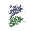
| ||||||||
|---|---|---|---|---|---|---|---|---|---|
| 1 | 
| ||||||||
| 2 | 
| ||||||||
| Unit cell |
|
-Components
| #1: Protein | Mass: 33241.965 Da / Num. of mol.: 2 / Fragment: residues 228-515 Source method: isolated from a genetically manipulated source Source: (gene. exp.)  References: UniProt: P00520, non-specific protein-tyrosine kinase #2: Chemical |   Mass: 372.420 Da / Num. of mol.: 2 / Source method: obtained synthetically / Formula: C22H20N4O2 Mass: 372.420 Da / Num. of mol.: 2 / Source method: obtained synthetically / Formula: C22H20N4O2#3: Water | ChemComp-HOH / |  Mass: 18.015 Da / Num. of mol.: 257 / Source method: isolated from a natural source / Formula: H2O Mass: 18.015 Da / Num. of mol.: 257 / Source method: isolated from a natural source / Formula: H2O |
|---|
-Experimental details
-Experiment
| Experiment | Method: X-RAY DIFFRACTION / Number of used crystals: 1 |
|---|
- Sample preparation
| Crystal | Density Matthews: 2.2 Å3/Da / Density % sol: 44.05 % |
|---|---|
| Crystal grow | Temperature: 277 K / Method: evaporation / pH: 8.5 Details: 30% PEG 4K, 0.1M Tris, 0.2M sodium acetate, pH 8.5, EVAPORATION, temperature 277K |
-Data collection
| Diffraction | Mean temperature: 100 K |
|---|---|
| Diffraction source | Source: SYNCHROTRON / Site: APS  / Beamline: 19-BM / Wavelength: 0.98 Å / Beamline: 19-BM / Wavelength: 0.98 Å |
| Detector | Type: ADSC QUANTUM 315 / Detector: CCD |
| Radiation | Protocol: SINGLE WAVELENGTH / Monochromatic (M) / Laue (L): M / Scattering type: x-ray |
| Radiation wavelength | Wavelength: 0.98 Å / Relative weight: 1 |
| Reflection | Resolution: 1.95→50 Å / Num. obs: 42995 / % possible obs: 93.8 % / Rmerge(I) obs: 0.083 / Net I/σ(I): 20 |
| Reflection shell | Resolution: 1.95→2.02 Å / Rmerge(I) obs: 0.324 / % possible all: 71 |
- Processing
| Software |
| ||||||||||||||||||
|---|---|---|---|---|---|---|---|---|---|---|---|---|---|---|---|---|---|---|---|
| Refinement | Method to determine structure: MOLECULAR REPLACEMENT Starting model: 2GQG Resolution: 1.95→50 Å / σ(F): 0 / σ(I): 0
| ||||||||||||||||||
| Refinement step | Cycle: LAST / Resolution: 1.95→50 Å
|
