 Movie
Movie Controller

[English] 日本語
 Yorodumi
- PDB-2qn4: Structure and function study of rice bifunctional alpha-amylase/s... -
+ Open data

- Basic information
| Entry | Database: PDB / ID: 2qn4 | ||||||
|---|---|---|---|---|---|---|---|
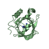
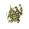
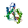
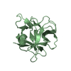
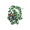
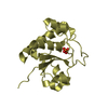
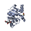
| Title | Structure and function study of rice bifunctional alpha-amylase/subtilisin inhibitor from Oryza sativa | ||||||
 Components Components | Alpha-amylase/subtilisin inhibitor | ||||||
 Keywords Keywords | HYDROLASE INHIBITOR / RASI / amylase inhibitor / subtilisin inhibitor / Alpha-amylase inhibitor / Protease inhibitor / Serine protease inhibitor | ||||||
| Function / homology |  Function and homology information Function and homology informationalpha-amylase inhibitor activity / serine-type endopeptidase inhibitor activity Similarity search - Function | ||||||
| Biological species |  | ||||||
| Method |  X-RAY DIFFRACTION / SYNCHROTRON / MOLECULAR REPLACEMENT / Resolution: 1.8 Å X-RAY DIFFRACTION / SYNCHROTRON / MOLECULAR REPLACEMENT / Resolution: 1.8 Å | ||||||
 Authors Authors | Peng, W.Y. / Lin, Y.H. / Huang, Y.C. / Guan, H.H. / Hsieh, Y.C. / Chen, C.J. | ||||||
 Citation Citation | Journal: To be Published Title: Structure and Function Study of Rice Bifunctional Alpha-Amylase/Subtilisin Inhibitor from Oryza Sativa Authors: Peng, W.Y. / Lin, Y.H. / Huang, Y.C. / Guan, H.H. / Hsieh, Y.C. / Liu, M.Y. / Chang, T. / Chen, C.J. #1: Journal: ACTA CRYSTALLOGR.,SECT.F / Year: 2006 Title: Purification, crystallization and preliminary X-ray crystallographic analysis of rice bifunctional alpha-amylase/subtilisin inhibitor from Oryza sativa Authors: Lin, Y.H. / Peng, W.Y. / Huang, Y.C. / Guan, H.H. / Hsieh, Y.C. / Liu, M.Y. / Chang, T. / Chen, C.J. | ||||||
| History |
|
- Structure visualization
| Structure viewer | Molecule: MolmilJmol/JSmol |
|---|
- Downloads & links
-Download
| PDBx/mmCIF format | 2qn4.cif.gz | 77.5 KB | Display | PDBx/mmCIF format |
|---|---|---|---|---|
| PDB format | pdb2qn4.ent.gz | 57.8 KB | Display | PDB format |
| PDBx/mmJSON format | 2qn4.json.gz | Tree view | PDBx/mmJSON format | |
| Others |  Other downloads Other downloads |
-Validation report
| Arichive directory | https://data.pdbj.org/pub/pdb/validation_reports/qn/2qn4ftp://data.pdbj.org/pub/pdb/validation_reports/qn/2qn4 | HTTPS FTP |
|---|
-Related structure data
| Related structure data | 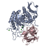 1avaS S: Starting model for refinement |
|---|---|
| Similar structure data |


-Links
 PDBj
PDBj- Assembly
| Deposited unit | 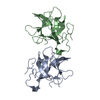
| ||||||||
|---|---|---|---|---|---|---|---|---|---|
| 1 | 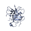
| ||||||||
| 2 | 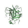
| ||||||||
| Unit cell |
|
-Components
| #1: Protein | Mass: 21443.461 Da / Num. of mol.: 2 / Source method: isolated from a natural source Source: (natural) Strain: japonica cultivar-group / References: UniProt: P29421 #2: Water | ChemComp-HOH / |  Mass: 18.015 Da / Num. of mol.: 198 / Source method: isolated from a natural source / Formula: H2O Mass: 18.015 Da / Num. of mol.: 198 / Source method: isolated from a natural source / Formula: H2OHas protein modification | Y | |
|---|
-Experimental details
-Experiment
| Experiment | Method: X-RAY DIFFRACTION / Number of used crystals: 1 |
|---|
- Sample preparation
| Crystal | Density Matthews: 1.96 Å3/Da / Density % sol: 37.17 % |
|---|---|
| Crystal grow | Temperature: 293 K / Method: vapor diffusion, hanging drop / pH: 4.7 Details: 20% PEG3350, 0.2M Potassium dihydrogen phosphate, pH4.7, VAPOR DIFFUSION, HANGING DROP, temperature 293K |
-Data collection
| Diffraction | Mean temperature: 100 K |
|---|---|
| Diffraction source | Source: SYNCHROTRON / Site: NSRRC  / Beamline: BL13B1 / Wavelength: 1 Å / Beamline: BL13B1 / Wavelength: 1 Å |
| Detector | Type: ADSC QUANTUM 315 / Detector: CCD / Date: Apr 6, 2006 Details: Vertically Collimating Premirror, LN2-Cooled Fixed-Exit Double Crystal Si(111) Monochromator, Toroidal Focusing Mirror |
| Radiation | Monochromator: LN2-Cooled, Fixed-Exit Double Crystal Monochromator Protocol: SINGLE WAVELENGTH / Monochromatic (M) / Laue (L): M / Scattering type: x-ray |
| Radiation wavelength | Wavelength: 1 Å / Relative weight: 1 |
| Reflection | Resolution: 1.8→30 Å / Num. obs: 31823 / % possible obs: 99.5 % / Redundancy: 4.7 % / Rsym value: 0.062 / Net I/σ(I): 22.5654 |
| Reflection shell | Resolution: 1.8→1.86 Å / Redundancy: 4.7 % / Rmerge(I) obs: 0.269 / Mean I/σ(I) obs: 5.25 / Num. unique all: 3129 / Rsym value: 0.363 / % possible all: 99.9 |
- Processing
| Software |
| |||||||||||||||||||||||||
|---|---|---|---|---|---|---|---|---|---|---|---|---|---|---|---|---|---|---|---|---|---|---|---|---|---|---|
| Refinement | Method to determine structure: MOLECULAR REPLACEMENT Starting model: PDB ENTRY 1AVA Resolution: 1.8→30 Å / Isotropic thermal model: Anisotropic / σ(F): 0 / σ(I): 0 / Stereochemistry target values: Engh & Huber
| |||||||||||||||||||||||||
| Displacement parameters | Biso mean: 0.16 Å2
| |||||||||||||||||||||||||
| Refine analyze |
| |||||||||||||||||||||||||
| Refinement step | Cycle: LAST / Resolution: 1.8→30 Å
| |||||||||||||||||||||||||
| Refine LS restraints |
| |||||||||||||||||||||||||
| LS refinement shell | Resolution: 1.8→1.86 Å
|
