- PDB-2qm3: Crystal structure of a predicted methyltransferase from Pyrococcu... -
+
Open data
ID or keywords:
Loading...
-
Basic information
Entry
Database: PDB / ID: 2qm3
Title
Crystal structure of a predicted methyltransferase from Pyrococcus furiosus
Components
Predicted methyltransferase
Keywords
TRANSFERASE / putative methyltransferase / structural genomics / Pyrococcus furiosus / PSI-2 / Protein Structure Initiative / Midwest Center for Structural Genomics / MCSG
Function / homology
Function and homology information
N4-bis(aminopropyl)spermidine synthase / polyamine biosynthetic process / transferase activity, transferring alkyl or aryl (other than methyl) groups / metal ion binding / cytoplasm Similarity search - Function
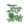
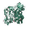
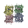
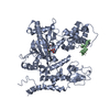
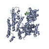
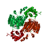
Authors state that the biological unit of this protein is experimentally unknown, and that the dimeric assembly shown in remark 350 is a prediction based on crystal packing.
-
Components
#1: Protein
Predictedmethyltransferase / Uncharacterized protein PF1111
Resolution: 2.05→39.9 Å / Num. all: 22125 / Num. obs: 22125 / % possible obs: 95.09 % / Observed criterion σ(I): -3 / Redundancy: 4.6 % / Biso Wilson estimate: 36.8 Å2 / Rmerge(I) obs: 0.078 / Net I/σ(I): 10
Reflection shell
Resolution: 2.05→2.1 Å / Redundancy: 3.4 % / Mean I/σ(I) obs: 2.2 / Rsym value: 0.415 / % possible all: 73.6
-
Processing
Software
Name
Version
Classification
REFMAC
5.2.0019
refinement
SBC-Collect
datacollection
HKL-3000
datareduction
HKL-3000
datascaling
HKL-3000
phasing
SHELXD
phasing
SHELXE
modelbuilding
MLPHARE
phasing
DM
phasing
SOLVE
phasing
RESOLVE
phasing
ARP/wARP
modelbuilding
CCP4
phasing
O
modelbuilding
Coot
modelbuilding
Refinement
Method to determine structure: MAD / Resolution: 2.05→39.9 Å / Cor.coef. Fo:Fc: 0.963 / Cor.coef. Fo:Fc free: 0.944 / SU B: 9.626 / SU ML: 0.133 / TLS residual ADP flag: LIKELY RESIDUAL / Cross valid method: THROUGHOUT / σ(F): 0 / ESU R: 0.215 / ESU R Free: 0.178 Stereochemistry target values: MAXIMUM LIKELIHOOD WITH PHASES Details: 1. The link (residues 88 through 99) between the N- and C-terminal structural domains is missing. For the two molecules related by the crystallographic 2-fold axis, the pairing of domains is ...Details: 1. The link (residues 88 through 99) between the N- and C-terminal structural domains is missing. For the two molecules related by the crystallographic 2-fold axis, the pairing of domains is ambiguous. The authors have modeled the more compact combination, but the other remains possible given the data at hand. 2. HYDROGENS HAVE BEEN ADDED IN THE RIDING POSITIONS.
Rfactor
Num. reflection
% reflection
Selection details
Rfree
0.22657
1189
5.1 %
RANDOM
Rwork
0.18296
-
-
-
obs
0.18518
22125
95.09 %
-
all
-
22125
-
-
Solvent computation
Ion probe radii: 0.8 Å / Shrinkage radii: 0.8 Å / VDW probe radii: 1.2 Å / Solvent model: MASK
Displacement parameters
Biso mean: 48.9 Å2
Baniso -1
Baniso -2
Baniso -3
1-
-2.14 Å2
0 Å2
0 Å2
2-
-
-1.84 Å2
0 Å2
3-
-
-
3.98 Å2
Refinement step
Cycle: LAST / Resolution: 2.05→39.9 Å
Protein
Nucleic acid
Ligand
Solvent
Total
Num. atoms
2770
0
14
223
3007
Refine LS restraints
Refine-ID
Type
Dev ideal
Dev ideal target
Number
X-RAY DIFFRACTION
r_bond_refined_d
0.014
0.022
2882
X-RAY DIFFRACTION
r_bond_other_d
X-RAY DIFFRACTION
r_angle_refined_deg
1.387
1.978
3910
X-RAY DIFFRACTION
r_angle_other_deg
X-RAY DIFFRACTION
r_dihedral_angle_1_deg
5.553
5
348
X-RAY DIFFRACTION
r_dihedral_angle_2_deg
36.845
24.138
145
X-RAY DIFFRACTION
r_dihedral_angle_3_deg
15.835
15
521
X-RAY DIFFRACTION
r_dihedral_angle_4_deg
22.952
15
22
X-RAY DIFFRACTION
r_chiral_restr
0.089
0.2
434
X-RAY DIFFRACTION
r_gen_planes_refined
0.006
0.02
2196
X-RAY DIFFRACTION
r_gen_planes_other
X-RAY DIFFRACTION
r_nbd_refined
0.213
0.2
1324
X-RAY DIFFRACTION
r_nbd_other
X-RAY DIFFRACTION
r_nbtor_refined
0.309
0.2
1997
X-RAY DIFFRACTION
r_nbtor_other
X-RAY DIFFRACTION
r_xyhbond_nbd_refined
0.165
0.2
216
X-RAY DIFFRACTION
r_xyhbond_nbd_other
X-RAY DIFFRACTION
r_metal_ion_refined
0.057
0.2
2
X-RAY DIFFRACTION
r_metal_ion_other
X-RAY DIFFRACTION
r_symmetry_vdw_refined
0.195
0.2
69
X-RAY DIFFRACTION
r_symmetry_vdw_other
X-RAY DIFFRACTION
r_symmetry_hbond_refined
0.137
0.2
19
X-RAY DIFFRACTION
r_symmetry_hbond_other
X-RAY DIFFRACTION
r_symmetry_metal_ion_refined
X-RAY DIFFRACTION
r_symmetry_metal_ion_other
X-RAY DIFFRACTION
r_mcbond_it
0.936
1.5
1758
X-RAY DIFFRACTION
r_mcbond_other
X-RAY DIFFRACTION
r_mcangle_it
1.405
2
2790
X-RAY DIFFRACTION
r_scbond_it
2.282
3
1268
X-RAY DIFFRACTION
r_scangle_it
3.517
4.5
1115
X-RAY DIFFRACTION
r_rigid_bond_restr
X-RAY DIFFRACTION
r_sphericity_free
X-RAY DIFFRACTION
r_sphericity_bonded
LS refinement shell
Resolution: 2.05→2.103 Å / Total num. of bins used: 20
Rfactor
Num. reflection
% reflection
Rfree
0.274
65
-
Rwork
0.233
1258
-
obs
-
-
74.16 %
Refinement TLS params.
Method: refined / Origin x: 47.9988 Å / Origin y: 12.366 Å / Origin z: 42.8343 Å
11
12
13
21
22
23
31
32
33
T
-0.0734 Å2
-0.0193 Å2
0.0102 Å2
-
-0.0967 Å2
-0.098 Å2
-
-
-0.1143 Å2
L
3.3019 °2
-0.4801 °2
0.0095 °2
-
1.3798 °2
-0.0348 °2
-
-
0.2605 °2
S
0.0489 Å °
-0.4184 Å °
0.5407 Å °
0.0955 Å °
-0.0205 Å °
-0.1954 Å °
-0.0725 Å °
0.0086 Å °
-0.0283 Å °
+
About Yorodumi
-
News
-
Feb 9, 2022. New format data for meta-information of EMDB entries
New format data for meta-information of EMDB entries
Version 3 of the EMDB header file is now the official format.
The previous official version 1.9 will be removed from the archive.
In the structure databanks used in Yorodumi, some data are registered as the other names, "COVID-19 virus" and "2019-nCoV". Here are the details of the virus and the list of structure data.
Jan 31, 2019. EMDB accession codes are about to change! (news from PDBe EMDB page)
EMDB accession codes are about to change! (news from PDBe EMDB page)
The allocation of 4 digits for EMDB accession codes will soon come to an end. Whilst these codes will remain in use, new EMDB accession codes will include an additional digit and will expand incrementally as the available range of codes is exhausted. The current 4-digit format prefixed with “EMD-” (i.e. EMD-XXXX) will advance to a 5-digit format (i.e. EMD-XXXXX), and so on. It is currently estimated that the 4-digit codes will be depleted around Spring 2019, at which point the 5-digit format will come into force.
The EM Navigator/Yorodumi systems omit the EMD- prefix.
Related info.:Q: What is EMD? / ID/Accession-code notation in Yorodumi/EM Navigator
Yorodumi is a browser for structure data from EMDB, PDB, SASBDB, etc.
This page is also the successor to EM Navigator detail page, and also detail information page/front-end page for Omokage search.
The word "yorodu" (or yorozu) is an old Japanese word meaning "ten thousand". "mi" (miru) is to see.
Related info.:EMDB / PDB / SASBDB / Comparison of 3 databanks / Yorodumi Search / Aug 31, 2016. New EM Navigator & Yorodumi / Yorodumi Papers / Jmol/JSmol / Function and homology information / Changes in new EM Navigator and Yorodumi
 Movie
Movie Controller

 Yorodumi
 Open data
 Basic information
 Components
 Keywords
 Function and homology information
 Pyrococcus furiosus DSM 3638 (archaea)
 X-RAY DIFFRACTION /
  Authors
 Citation
 Structure visualization
 Downloads & links
 Other downloads


 PDBj
PDBj

 Assembly


 Mass: 40.078 Da / Num. of mol.: 2 / Source method: obtained synthetically / Formula: Ca
 Mass: 60.052 Da / Num. of mol.: 3 / Source method: obtained synthetically / Formula: C2H4O2
 Mass: 18.015 Da / Num. of mol.: 223 / Source method: isolated from a natural source / Formula: H2O
 Sample preparation
 / Beamline: 19-ID / Wavelength: 0.97918, 0.97932
 Processing