 Movie
Movie Controller


+ Open data

- Basic information
| Entry | Database: PDB / ID: 2qks | ||||||
|---|---|---|---|---|---|---|---|
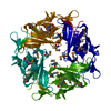
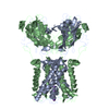
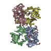
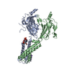
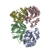
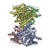
| Title | Crystal structure of a Kir3.1-prokaryotic Kir channel chimera | ||||||
 Components Components | Kir3.1-prokaryotic Kir channel chimera | ||||||
 Keywords Keywords | METAL TRANSPORT / CHIMERA / G-PROTEIN GATED INWARD RECTIFIER / POTASSIUM CHANNEL / SELECTIVITY FILTER | ||||||
| Function / homology |  Function and homology information Function and homology informationI(KACh) inward rectifier potassium channel complex / G-protein activated inward rectifier potassium channel activity / Activation of G protein gated Potassium channels / Inhibition of voltage gated Ca2+ channels via Gbeta/gamma subunits / inward rectifier potassium channel complex / voltage-gated monoatomic ion channel activity involved in regulation of presynaptic membrane potential / inward rectifier potassium channel activity / regulation of monoatomic ion transmembrane transport / potassium ion import across plasma membrane / parallel fiber to Purkinje cell synapse ...I(KACh) inward rectifier potassium channel complex / G-protein activated inward rectifier potassium channel activity / Activation of G protein gated Potassium channels / Inhibition of voltage gated Ca2+ channels via Gbeta/gamma subunits / inward rectifier potassium channel complex / voltage-gated monoatomic ion channel activity involved in regulation of presynaptic membrane potential / inward rectifier potassium channel activity / regulation of monoatomic ion transmembrane transport / potassium ion import across plasma membrane / parallel fiber to Purkinje cell synapse / monoatomic ion channel complex / response to electrical stimulus / phosphatidylinositol-4,5-bisphosphate binding / voltage-gated potassium channel complex / T-tubule / potassium ion transmembrane transport / presynaptic membrane / external side of plasma membrane / cell surface / plasma membrane Similarity search - Function | ||||||
| Biological species |   Paraburkholderia xenovorans (bacteria) Paraburkholderia xenovorans (bacteria) | ||||||
| Method |  X-RAY DIFFRACTION / SYNCHROTRON / MOLECULAR REPLACEMENT / Resolution: 2.2 Å X-RAY DIFFRACTION / SYNCHROTRON / MOLECULAR REPLACEMENT / Resolution: 2.2 Å | ||||||
 Authors Authors | Nishida, M. / MacKinnon, R. | ||||||
 Citation Citation | Journal: Embo J. / Year: 2007 Title: Crystal structure of a Kir3.1-prokaryotic Kir channel chimera. Authors: Nishida, M. / Cadene, M. / Chait, B.T. / Mackinnon, R. | ||||||
| History |
|
- Structure visualization
| Structure viewer | Molecule: MolmilJmol/JSmol |
|---|
- Downloads & links
-Download
| PDBx/mmCIF format | 2qks.cif.gz | 142.1 KB | Display | PDBx/mmCIF format |
|---|---|---|---|---|
| PDB format | pdb2qks.ent.gz | 106.5 KB | Display | PDB format |
| PDBx/mmJSON format | 2qks.json.gz | Tree view | PDBx/mmJSON format | |
| Others |  Other downloads Other downloads |
-Validation report
| Arichive directory | https://data.pdbj.org/pub/pdb/validation_reports/qk/2qksftp://data.pdbj.org/pub/pdb/validation_reports/qk/2qks | HTTPS FTP |
|---|
-Related structure data
-Links
 PDBj
PDBj


- Assembly
| Deposited unit | 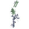
| |||||||||||||||||||||||||||||||||||||||||||||||||||
|---|---|---|---|---|---|---|---|---|---|---|---|---|---|---|---|---|---|---|---|---|---|---|---|---|---|---|---|---|---|---|---|---|---|---|---|---|---|---|---|---|---|---|---|---|---|---|---|---|---|---|---|---|
| 1 | 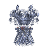
| |||||||||||||||||||||||||||||||||||||||||||||||||||
| 2 | 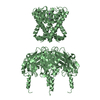
| |||||||||||||||||||||||||||||||||||||||||||||||||||
| Unit cell |
| |||||||||||||||||||||||||||||||||||||||||||||||||||
| Components on special symmetry positions |
| |||||||||||||||||||||||||||||||||||||||||||||||||||
| Details | The biological tetrameric assembly of the chain A molecule is generated by the operations: (y, 1-x, z), (1-x, 1-y, z) and (1-y, x, z). The biological tetrameric assembly of the chain B molecule is generated by the operations: (1-y, -1+x, z), (2-x, -y, z) and (1+y, 1-x, z). |
-Components
| #1: Protein | Mass: 36176.582 Da / Num. of mol.: 2 Fragment: KIR3.1 (residues 3-44, 125-318), KIRBAC1.3 TRANSMEMBRANE domain (residues 45-124) Mutation: M127A Source method: isolated from a genetically manipulated source Source: (gene. exp.) Paraburkholderia xenovorans (bacteria)Genus: Burkholderia / Plasmid: pET28b(+) / Strain: LB400 / Species (production host): Escherichia coli / Production host: #2: Sugar | ChemComp-BNG / |   Type: D-saccharide / Mass: 306.395 Da / Num. of mol.: 1 Type: D-saccharide / Mass: 306.395 Da / Num. of mol.: 1Source method: isolated from a genetically manipulated source Formula: C15H30O6 / Comment: detergent*YM #3: Chemical | ChemComp-K /   Mass: 39.098 Da / Num. of mol.: 12 / Source method: obtained synthetically / Formula: K Mass: 39.098 Da / Num. of mol.: 12 / Source method: obtained synthetically / Formula: K#4: Water | ChemComp-HOH / |  Mass: 18.015 Da / Num. of mol.: 226 / Source method: isolated from a natural source / Formula: H2O Mass: 18.015 Da / Num. of mol.: 226 / Source method: isolated from a natural source / Formula: H2O |
|---|
-Experimental details
-Experiment
| Experiment | Method: X-RAY DIFFRACTION / Number of used crystals: 1 |
|---|
- Sample preparation
| Crystal | Density Matthews: 3.1 Å3/Da / Density % sol: 60.3 % |
|---|---|
| Crystal grow | Temperature: 293 K / Method: vapor diffusion, sitting drop / pH: 6.2 Details: 10-15% (w/v) PEG 4000, 0.05M sodium citrate, 0.2M potassium phosphate, 0.08M bis-tris, 0.12M KCl, 0.003M dithiothreitol, 0.02M tris (2-carboxyethyl) phosphine hydrochloride, 0.0026M 1,2- ...Details: 10-15% (w/v) PEG 4000, 0.05M sodium citrate, 0.2M potassium phosphate, 0.08M bis-tris, 0.12M KCl, 0.003M dithiothreitol, 0.02M tris (2-carboxyethyl) phosphine hydrochloride, 0.0026M 1,2-dihexanoyl phosphatidylinositol 4,5-biphosphate, 0.016M nonylglucoside , pH 6.2, VAPOR DIFFUSION, SITTING DROP, temperature 293.0K |
-Data collection
| Diffraction | Mean temperature: 100 K |
|---|---|
| Diffraction source | Source: SYNCHROTRON / Site: NSLS  / Beamline: X29A / Wavelength: 1.1 Å / Beamline: X29A / Wavelength: 1.1 Å |
| Detector | Type: ADSC QUANTUM 315 / Detector: CCD / Date: Aug 2, 2006 |
| Radiation | Monochromator: DOUBLE CRYSTALS (Silicon) / Protocol: SINGLE WAVELENGTH / Monochromatic (M) / Laue (L): M / Scattering type: x-ray |
| Radiation wavelength | Wavelength: 1.1 Å / Relative weight: 1 |
| Reflection | Resolution: 2.2→100 Å / Num. all: 43991 / Num. obs: 43991 / % possible obs: 97.9 % / Observed criterion σ(F): 0 / Observed criterion σ(I): -3 / Redundancy: 3.7 % / Biso Wilson estimate: 46.1 Å2 / Rsym value: 0.054 / Net I/σ(I): 17 |
| Reflection shell | Resolution: 2.2→2.28 Å / Redundancy: 3.7 % / Mean I/σ(I) obs: 3.9 / Num. unique all: 4450 / Rsym value: 0.309 / % possible all: 99.7 |
- Processing
| Software |
| ||||||||||||||||||||||||||||
|---|---|---|---|---|---|---|---|---|---|---|---|---|---|---|---|---|---|---|---|---|---|---|---|---|---|---|---|---|---|
| Refinement | Method to determine structure: MOLECULAR REPLACEMENT Starting model: PDB ENTRIES 1N9P, 1P7B Resolution: 2.2→29.5 Å Isotropic thermal model: Anisotropic model for the reflection data and restrained individual isotoropic temperature factors for the coordinates Cross valid method: THROUGHOUT / σ(F): 0 / Stereochemistry target values: Engh & Huber Details: During the refinement, only the anisotropic components of the correction were applied to the data, the isotropic component was not applied to the coordinates. The bulk solvent correction was ...Details: During the refinement, only the anisotropic components of the correction were applied to the data, the isotropic component was not applied to the coordinates. The bulk solvent correction was applied between 5-2.2 A. Potassium ions are located using another crystal soaked in a rubidium-containing solution.
| ||||||||||||||||||||||||||||
| Displacement parameters | Biso mean: 51.229 Å2
| ||||||||||||||||||||||||||||
| Refine analyze |
| ||||||||||||||||||||||||||||
| Refinement step | Cycle: LAST / Resolution: 2.2→29.5 Å
| ||||||||||||||||||||||||||||
| Refine LS restraints |
| ||||||||||||||||||||||||||||
| LS refinement shell | Resolution: 2.2→2.37 Å / Rfactor Rfree error: 0.015
|
