 Movie
Movie Controller

[English] 日本語
 Yorodumi
- PDB-7kaz: Crystal structure of OhyA(E82A)-18:1/h18:0-FAD complex from Staph... -
+ Open data

- Basic information
| Entry | Database: PDB / ID: 7kaz | |||||||||
|---|---|---|---|---|---|---|---|---|---|---|
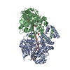
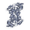
| Title | Crystal structure of OhyA(E82A)-18:1/h18:0-FAD complex from Staphylococcus aureus | |||||||||
 Components Components | Oleate hydratase | |||||||||
 Keywords Keywords | OXIDOREDUCTASE / Oleate hydratase / OhyA / SaOhyA / 18:1 / complex / oleic acid / hydroxystearic acid / FAD / h18:0 | |||||||||
| Function / homology |  Function and homology information Function and homology informationoleate hydratase / oleate hydratase activity / FAD binding / fatty acid metabolic process Similarity search - Function | |||||||||
| Biological species |   Staphylococcus aureus (bacteria) Staphylococcus aureus (bacteria) | |||||||||
| Method |  X-RAY DIFFRACTION / SYNCHROTRON / MOLECULAR REPLACEMENT / Resolution: 1.854 Å X-RAY DIFFRACTION / SYNCHROTRON / MOLECULAR REPLACEMENT / Resolution: 1.854 Å | |||||||||
 Authors Authors | Radka, C.D. / Rock, C.O. | |||||||||
| Funding support |  United States, 2items United States, 2items
| |||||||||
 Citation Citation | Journal: J.Biol.Chem. / Year: 2021 Title: Structure and mechanism of Staphylococcus aureus oleate hydratase (OhyA). Authors: Radka, C.D. / Batte, J.L. / Frank, M.W. / Young, B.M. / Rock, C.O. | |||||||||
| History |
|
- Structure visualization
| Structure viewer | Molecule: MolmilJmol/JSmol |
|---|
- Downloads & links
-Download
| PDBx/mmCIF format | 7kaz.cif.gz | 731.7 KB | Display | PDBx/mmCIF format |
|---|---|---|---|---|
| PDB format | pdb7kaz.ent.gz | 598.7 KB | Display | PDB format |
| PDBx/mmJSON format | 7kaz.json.gz | Tree view | PDBx/mmJSON format | |
| Others |  Other downloads Other downloads |
-Validation report
| Arichive directory | https://data.pdbj.org/pub/pdb/validation_reports/ka/7kazftp://data.pdbj.org/pub/pdb/validation_reports/ka/7kaz | HTTPS FTP |
|---|
-Related structure data
| Related structure data |  7kavSC  7kawC  7kaxC  7kayC S: Starting model for refinement C: citing same article ( |
|---|---|
| Similar structure data |


-Links
 PDBj
PDBj


- Assembly
| Deposited unit | 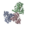
| |||||||||||||||
|---|---|---|---|---|---|---|---|---|---|---|---|---|---|---|---|---|
| 1 | 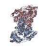
| |||||||||||||||
| 2 | 
| |||||||||||||||
| Unit cell |
| |||||||||||||||
| Components on special symmetry positions |
|
-Components
-Protein , 1 types, 3 molecules CBA
| #1: Protein | Mass: 69834.688 Da / Num. of mol.: 3 / Mutation: E82A Source method: isolated from a genetically manipulated source Source: (gene. exp.) Staphylococcus aureus (bacteria)Gene: DD547_00094, DQV53_00770, EP54_06595, EQ90_12415, G0V24_00735, G0X12_00605, G0Z18_00840, G6Y10_10285, GO746_00220, GO803_11440, GO805_08895, GO821_10135, GO894_07145, GO942_14045, HMPREF3211_ ...Gene: DD547_00094, DQV53_00770, EP54_06595, EQ90_12415, G0V24_00735, G0X12_00605, G0Z18_00840, G6Y10_10285, GO746_00220, GO803_11440, GO805_08895, GO821_10135, GO894_07145, GO942_14045, HMPREF3211_02399, NCTC10654_00136, RK64_00980 Production host: |
|---|
-Non-polymers , 5 types, 2731 molecules 


| #2: Chemical | ChemComp-WAD / ( Mass: 300.477 Da / Num. of mol.: 1 / Source method: obtained synthetically / Formula: C18H36O3 / Feature type: SUBJECT OF INVESTIGATION Mass: 300.477 Da / Num. of mol.: 1 / Source method: obtained synthetically / Formula: C18H36O3 / Feature type: SUBJECT OF INVESTIGATION | ||||||
|---|---|---|---|---|---|---|---|
| #3: Chemical |  Mass: 785.550 Da / Num. of mol.: 2 / Source method: obtained synthetically / Formula: C27H33N9O15P2 / Feature type: SUBJECT OF INVESTIGATION / Comment: FAD*YM Mass: 785.550 Da / Num. of mol.: 2 / Source method: obtained synthetically / Formula: C27H33N9O15P2 / Feature type: SUBJECT OF INVESTIGATION / Comment: FAD*YM#4: Chemical | ChemComp-K / |  Mass: 39.098 Da / Num. of mol.: 1 / Source method: obtained synthetically / Formula: K Mass: 39.098 Da / Num. of mol.: 1 / Source method: obtained synthetically / Formula: K#5: Chemical |  Mass: 282.461 Da / Num. of mol.: 2 / Source method: obtained synthetically / Formula: C18H34O2 / Feature type: SUBJECT OF INVESTIGATION Mass: 282.461 Da / Num. of mol.: 2 / Source method: obtained synthetically / Formula: C18H34O2 / Feature type: SUBJECT OF INVESTIGATION#6: Water | ChemComp-HOH / | Mass: 18.015 Da / Num. of mol.: 2725 / Source method: isolated from a natural source / Formula: H2O |
-Details
| Has ligand of interest | Y |
|---|
-Experimental details
-Experiment
| Experiment | Method: X-RAY DIFFRACTION / Number of used crystals: 1 |
|---|
- Sample preparation
| Crystal | Density Matthews: 2.68 Å3/Da / Density % sol: 54.08 % |
|---|---|
| Crystal grow | Temperature: 293.15 K / Method: vapor diffusion, hanging drop / pH: 6.5 Details: Crystallized: 15% v/v PEG 3000, 200 mM magnesium chloride, 100 mM sodium cacodylate, pH 6.5; Crystals soaked in 15% PEG3000, 200 mM magnesium chloride, 100 mM sodium cacodylate, pH 6.5, and ...Details: Crystallized: 15% v/v PEG 3000, 200 mM magnesium chloride, 100 mM sodium cacodylate, pH 6.5; Crystals soaked in 15% PEG3000, 200 mM magnesium chloride, 100 mM sodium cacodylate, pH 6.5, and 750 uM FAD. Cryo: 15% v/v PEG3000, 200 mM magnesium chloride, 100 mM sodium cacodylate, pH 6.5, 30% glycerol |
-Data collection
| Diffraction | Mean temperature: 100 K / Serial crystal experiment: N |
|---|---|
| Diffraction source | Source: SYNCHROTRON / Site: APS / Beamline: 22-ID / Wavelength: 1 Å |
| Detector | Type: DECTRIS EIGER X 16M / Detector: PIXEL / Date: Nov 8, 2019 |
| Radiation | Monochromator: Si 111. Rosenbaum-Rock double-crystal monochromator: liquid nitrogen cooled; sagitally focusing 2nd crystal, Rosenbaum-Rock vertical focusing mirror Protocol: SINGLE WAVELENGTH / Monochromatic (M) / Laue (L): M / Scattering type: x-ray |
| Radiation wavelength | Wavelength: 1 Å / Relative weight: 1 |
| Reflection | Resolution: 1.85→22.42 Å / Num. obs: 183821 / % possible obs: 98.5 % / Redundancy: 3.8 % / Rpim(I) all: 0.04 / Net I/σ(I): 6.4 |
| Reflection shell | Resolution: 1.85→1.9 Å / Redundancy: 3.8 % / Num. unique obs: 18078 / Rpim(I) all: 0.485 / % possible all: 96.3 |
- Processing
| Software |
| ||||||||||||||||||||||||||||||||||||||||||||||||||||||||||||||||||||||||||||||||||||||||||
|---|---|---|---|---|---|---|---|---|---|---|---|---|---|---|---|---|---|---|---|---|---|---|---|---|---|---|---|---|---|---|---|---|---|---|---|---|---|---|---|---|---|---|---|---|---|---|---|---|---|---|---|---|---|---|---|---|---|---|---|---|---|---|---|---|---|---|---|---|---|---|---|---|---|---|---|---|---|---|---|---|---|---|---|---|---|---|---|---|---|---|---|
| Refinement | Method to determine structure: MOLECULAR REPLACEMENT Starting model: 7KAV Resolution: 1.854→22.42 Å / SU ML: 0.33 / Cross valid method: THROUGHOUT / σ(F): 1.42 / Phase error: 27.77 / Stereochemistry target values: ML
| ||||||||||||||||||||||||||||||||||||||||||||||||||||||||||||||||||||||||||||||||||||||||||
| Solvent computation | Shrinkage radii: 0.9 Å / VDW probe radii: 1.11 Å / Solvent model: FLAT BULK SOLVENT MODEL | ||||||||||||||||||||||||||||||||||||||||||||||||||||||||||||||||||||||||||||||||||||||||||
| Displacement parameters | Biso max: 112.29 Å2 / Biso mean: 29.2785 Å2 / Biso min: 6.04 Å2 | ||||||||||||||||||||||||||||||||||||||||||||||||||||||||||||||||||||||||||||||||||||||||||
| Refinement step | Cycle: final / Resolution: 1.854→22.42 Å
| ||||||||||||||||||||||||||||||||||||||||||||||||||||||||||||||||||||||||||||||||||||||||||
| Refine LS restraints |
| ||||||||||||||||||||||||||||||||||||||||||||||||||||||||||||||||||||||||||||||||||||||||||
| LS refinement shell | Refine-ID: X-RAY DIFFRACTION / Rfactor Rfree error: 0
|
