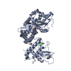
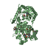
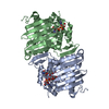
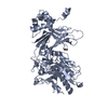
- PDB-2qj8: Crystal structure of an aspartoacylase family protein (mlr6093) f... -
+
Open data
ID or keywords:
Loading...
-
Basic information
Entry
Database: PDB / ID: 2qj8
Title
Crystal structure of an aspartoacylase family protein (mlr6093) from mesorhizobium loti maff303099 at 2.00 A resolution
Components
Mlr6093 protein
Keywords
HYDROLASE / Structural genomics / Joint Center for Structural Genomics / JCSG / Protein Structure Initiative / PSI-2
Function / homology
Function and homology information
hydrolase activity, acting on carbon-nitrogen (but not peptide) bonds, in linear amides / hydrolase activity, acting on ester bonds / metal ion binding Similarity search - Function
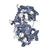
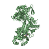
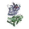
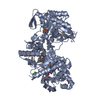
BIOMOLECULE: 1 THIS ENTRY CONTAINS THE CRYSTALLOGRAPHIC ASYMMETRIC UNIT WHICH CONSISTS OF 2 CHAINS ... BIOMOLECULE: 1 THIS ENTRY CONTAINS THE CRYSTALLOGRAPHIC ASYMMETRIC UNIT WHICH CONSISTS OF 2 CHAINS WHICH FORM A DIMER BASED ON CRYSTAL PACKING ANALYSIS. SIZE EXCLUSION CHROMATOGRAPHY SUPPORTS THE ASSIGNMENT OF A DIMER AS THE SIGNIFICANT OLIGOMERIZATION STATE. SEE REMARK 350 FOR INFORMATION ON GENERATING THE BIOLOGICAL MOLECULE(S).
Remark 999
SEQUENCE THE CONSTRUCT WAS EXPRESSED WITH A PURIFICATION TAG MGSDKIHHHHHHENLYFQG. THE TAG WAS ... SEQUENCE THE CONSTRUCT WAS EXPRESSED WITH A PURIFICATION TAG MGSDKIHHHHHHENLYFQG. THE TAG WAS REMOVED WITH TEV PROTEASE LEAVING ONLY A GLYCINE FOLLOWED BY THE TARGET SEQUENCE.
Type: ADSC QUANTUM 4 / Detector: CCD / Date: Jun 9, 2007 Details: 1m long Rh coated bent cylindrical mirror for horizontal and vertical focusing
Radiation
Monochromator: Double-crystal / Protocol: SINGLE WAVELENGTH / Monochromatic (M) / Laue (L): M / Scattering type: x-ray
Radiation wavelength
Wavelength: 0.978662 Å / Relative weight: 1
Reflection
Resolution: 2→29.643 Å / Num. obs: 45402 / % possible obs: 99.8 % / Redundancy: 4.6 % / Biso Wilson estimate: 26.79 Å2 / Rmerge(I) obs: 0.105 / Rsym value: 0.105 / Net I/σ(I): 11.5
Reflection shell
Diffraction-ID: 1
Resolution (Å)
Redundancy (%)
Rmerge(I) obs
Mean I/σ(I) obs
Num. measured all
Num. unique all
Rsym value
% possible all
2-2.05
3.8
0.782
1.7
12285
3268
0.782
98.7
2.05-2.11
4.6
0.744
0.6
15029
3241
0.744
100
2.11-2.17
4.8
0.553
1.4
15018
3135
0.553
100
2.17-2.24
4.7
0.499
1.1
14235
3061
0.499
100
2.24-2.31
4.7
0.43
1
13985
2968
0.43
100
2.31-2.39
4.8
0.316
2.4
13709
2864
0.316
100
2.39-2.48
4.8
0.267
1.9
13382
2790
0.267
100
2.48-2.58
4.8
0.211
3.5
12768
2670
0.211
100
2.58-2.7
4.7
0.19
3.4
12094
2584
0.19
100
2.7-2.83
4.8
0.141
5.2
11679
2446
0.141
100
2.83-2.98
4.8
0.114
6.2
11202
2354
0.114
100
2.98-3.16
4.8
0.092
7.5
10605
2219
0.092
100
3.16-3.38
4.7
0.074
9
9965
2122
0.074
100
3.38-3.65
4.6
0.068
8.2
8897
1932
0.068
100
3.65-4
4.6
0.059
10
8325
1827
0.059
100
4-4.47
4.7
0.048
12.6
7713
1652
0.048
100
4.47-5.16
4.6
0.051
11.6
6745
1464
0.051
100
5.16-6.32
4.5
0.058
10.3
5636
1256
0.058
99.8
6.32-8.94
4.3
0.047
13.1
4320
994
0.047
99.6
8.94-29.643
3.9
0.044
13.4
2158
555
0.044
95.9
-
Phasing
Phasing
Method: SAD
-
Processing
Software
Name
Version
Classification
NB
REFMAC
5.2.0019
refinement
PHENIX
refinement
SHELX
phasing
MolProbity
3beta29
modelbuilding
SCALA
datascaling
PDB_EXTRACT
3
dataextraction
ADSC
Quantum
datacollection
MOSFLM
datareduction
SHELXD
phasing
autoSHARP
phasing
Refinement
Method to determine structure: SAD / Resolution: 2→29.643 Å / Cor.coef. Fo:Fc: 0.947 / Cor.coef. Fo:Fc free: 0.921 / SU B: 10.618 / SU ML: 0.146 / TLS residual ADP flag: LIKELY RESIDUAL / Cross valid method: THROUGHOUT / σ(F): 0 / ESU R: 0.194 / ESU R Free: 0.175 Stereochemistry target values: MAXIMUM LIKELIHOOD WITH PHASES Details: 1. HYDROGENS HAVE BEEN ADDED IN THE RIDING POSITIONS. 2. A MET-INHIBITION PROTOCOL WAS USED FOR SELENOMETHIONINE INCORPORATION DURING PROTEIN EXPRESSION. THE OCCUPANCY OF THE SE ATOMS IN THE ...Details: 1. HYDROGENS HAVE BEEN ADDED IN THE RIDING POSITIONS. 2. A MET-INHIBITION PROTOCOL WAS USED FOR SELENOMETHIONINE INCORPORATION DURING PROTEIN EXPRESSION. THE OCCUPANCY OF THE SE ATOMS IN THE MSE RESIDUES WAS REDUCED TO 0.75 TO ACCOUNT FOR THE REDUCED SCATTERING POWER DUE TO PARTIAL S-MET INCORPORATION. 3. ATOM RECORD CONTAINS RESIDUAL B FACTORS ONLY. 4. RESIDUES A210-A215, A238-A247, B26-B28, B91-B115, B156-B160, B183-B188 AND B213-B214 ARE DISORDERED AND HAVE NOT BEEN MODELLED.
Rfactor
Num. reflection
% reflection
Selection details
Rfree
0.254
2286
5 %
RANDOM
Rwork
0.207
-
-
-
obs
0.209
45330
99.77 %
-
Solvent computation
Ion probe radii: 0.8 Å / Shrinkage radii: 0.8 Å / VDW probe radii: 1.2 Å / Solvent model: MASK
In the structure databanks used in Yorodumi, some data are registered as the other names, "COVID-19 virus" and "2019-nCoV". Here are the details of the virus and the list of structure data.
Jan 31, 2019. EMDB accession codes are about to change! (news from PDBe EMDB page)
EMDB accession codes are about to change! (news from PDBe EMDB page)
The allocation of 4 digits for EMDB accession codes will soon come to an end. Whilst these codes will remain in use, new EMDB accession codes will include an additional digit and will expand incrementally as the available range of codes is exhausted. The current 4-digit format prefixed with “EMD-” (i.e. EMD-XXXX) will advance to a 5-digit format (i.e. EMD-XXXXX), and so on. It is currently estimated that the 4-digit codes will be depleted around Spring 2019, at which point the 5-digit format will come into force.
The EM Navigator/Yorodumi systems omit the EMD- prefix.
Related info.:Q: What is EMD? / ID/Accession-code notation in Yorodumi/EM Navigator
Yorodumi is a browser for structure data from EMDB, PDB, SASBDB, etc.
This page is also the successor to EM Navigator detail page, and also detail information page/front-end page for Omokage search.
The word "yorodu" (or yorozu) is an old Japanese word meaning "ten thousand". "mi" (miru) is to see.
Related info.:EMDB / PDB / SASBDB / Comparison of 3 databanks / Yorodumi Search / Aug 31, 2016. New EM Navigator & Yorodumi / Yorodumi Papers / Jmol/JSmol / Function and homology information / Changes in new EM Navigator and Yorodumi
 Movie
Movie Controller

 Yorodumi
 Open data
 Basic information
 Components
 Keywords
 Function and homology information
 Mesorhizobium loti (bacteria)
 X-RAY DIFFRACTION /
  Authors
 Citation
 Structure visualization
 Downloads & links
 Other downloads
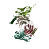

 PDBj
PDBj Assembly
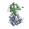
 Mass: 18.015 Da / Num. of mol.: 282 / Source method: isolated from a natural source / Formula: H2O
 Sample preparation
 / Beamline: BL1-5 / Wavelength: 0.978662 Å
 Processing