 Movie
Movie Controller

[English] 日本語
 Yorodumi
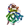
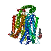
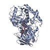
- PDB-2qgi: The UDP complex structure of the sixth gene product of the F1-ATP... -
+ Open data

- Basic information
| Entry | Database: PDB / ID: 2qgi | ||||||
|---|---|---|---|---|---|---|---|
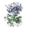
| Title | The UDP complex structure of the sixth gene product of the F1-ATPase operon of Rhodobacter blasticus | ||||||
 Components Components | ATP synthase subunits region ORF 6 | ||||||
 Keywords Keywords | TRANSFERASE / MAJASTRIDIN / ATPASE OPERON / GLYCOSYL TRANSFERASE / ROSSMANN FOLD / UDP-COMPLEX | ||||||
| Function / homology | Spore Coat Polysaccharide Biosynthesis Protein SpsA; Chain A / Spore Coat Polysaccharide Biosynthesis Protein SpsA; Chain A / Nucleotide-diphospho-sugar transferases / Alpha-Beta Complex / Alpha Beta / : / URIDINE-5'-DIPHOSPHATE / ATP synthase subunits region ORF 6 Function and homology information Function and homology information | ||||||
| Biological species |  Rhodobacter blasticus (bacteria) Rhodobacter blasticus (bacteria) | ||||||
| Method |  X-RAY DIFFRACTION / SYNCHROTRON / MOLECULAR REPLACEMENT / Resolution: 1.65 Å X-RAY DIFFRACTION / SYNCHROTRON / MOLECULAR REPLACEMENT / Resolution: 1.65 Å | ||||||
 Authors Authors | Enroth, C. / Strid, A. | ||||||
 Citation Citation | Journal: Biochim.Biophys.Acta / Year: 2008 Title: Crystal structure of a protein, structurally related to glycosyltransferases, encoded in the Rhodobacter blasticus atp operon. Authors: Enroth, C. / Strid, A. | ||||||
| History |
|
- Structure visualization
| Structure viewer | Molecule: MolmilJmol/JSmol |
|---|
- Downloads & links
-Download
| PDBx/mmCIF format | 2qgi.cif.gz | 133.4 KB | Display | PDBx/mmCIF format |
|---|---|---|---|---|
| PDB format | pdb2qgi.ent.gz | 100.4 KB | Display | PDB format |
| PDBx/mmJSON format | 2qgi.json.gz | Tree view | PDBx/mmJSON format | |
| Others |  Other downloads Other downloads |
-Validation report
| Arichive directory | https://data.pdbj.org/pub/pdb/validation_reports/qg/2qgiftp://data.pdbj.org/pub/pdb/validation_reports/qg/2qgi | HTTPS FTP |
|---|
-Related structure data
| Related structure data | 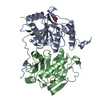 2nxvSC S: Starting model for refinement C: citing same article ( |
|---|---|
| Similar structure data |


-Links
 PDBj
PDBj


- Assembly
| Deposited unit | 
| ||||||||||||
|---|---|---|---|---|---|---|---|---|---|---|---|---|---|
| 1 |
| ||||||||||||
| 2 | 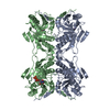
| ||||||||||||
| Unit cell |
| ||||||||||||
| Components on special symmetry positions |
| ||||||||||||
| Details | The assymmetric unit contains one biological unit, i.e. a dimer. |
-Components
| #1: Protein | Mass: 28720.590 Da / Num. of mol.: 2 Source method: isolated from a genetically manipulated source Source: (gene. exp.) Rhodobacter blasticus (bacteria) / Production host: #2: Chemical |   Mass: 54.938 Da / Num. of mol.: 2 / Source method: obtained synthetically / Formula: Mn Mass: 54.938 Da / Num. of mol.: 2 / Source method: obtained synthetically / Formula: Mn#3: Chemical | ChemComp-UDP / |   Type: RNA linking / Mass: 404.161 Da / Num. of mol.: 1 / Source method: obtained synthetically / Formula: C9H14N2O12P2 / Comment: UDP*YM Type: RNA linking / Mass: 404.161 Da / Num. of mol.: 1 / Source method: obtained synthetically / Formula: C9H14N2O12P2 / Comment: UDP*YM#4: Water | ChemComp-HOH / |  Mass: 18.015 Da / Num. of mol.: 721 / Source method: isolated from a natural source / Formula: H2O Mass: 18.015 Da / Num. of mol.: 721 / Source method: isolated from a natural source / Formula: H2O |
|---|
-Experimental details
-Experiment
| Experiment | Method: X-RAY DIFFRACTION / Number of used crystals: 1 |
|---|
- Sample preparation
| Crystal | Density Matthews: 3.36 Å3/Da / Density % sol: 63.37 % |
|---|---|
| Crystal grow | Temperature: 298 K / Method: vapor diffusion, hanging drop / pH: 6 Details: 100mM sodium citrate (pH6.0) and 75mM magnesium acetate. Mixed 4+4ul with protein sample, hanging drop, vapor diffusion, temperature 298K., VAPOR DIFFUSION, HANGING DROP |
-Data collection
| Diffraction | Mean temperature: 100 K |
|---|---|
| Diffraction source | Source: SYNCHROTRON / Site: MAX II  / Beamline: I911-2 / Wavelength: 1.043 Å / Beamline: I911-2 / Wavelength: 1.043 Å |
| Detector | Type: MAR CCD 165 mm / Detector: CCD / Date: May 1, 2006 |
| Radiation | Protocol: SINGLE WAVELENGTH / Monochromatic (M) / Laue (L): M / Scattering type: x-ray |
| Radiation wavelength | Wavelength: 1.043 Å / Relative weight: 1 |
| Reflection | Resolution: 1.65→20 Å / Num. obs: 93646 / % possible obs: 99.9 % / Redundancy: 6.1 % / Biso Wilson estimate: 18.1 Å2 / Rsym value: 0.071 / Net I/σ(I): 16.7 |
| Reflection shell | Resolution: 1.65→1.74 Å / Redundancy: 6.1 % / Mean I/σ(I) obs: 4.6 / Num. unique all: 13554 / Rsym value: 0.388 / % possible all: 100 |
- Processing
| Software |
| ||||||||||||||||||||||||||||||||||||||||||||||||||||||||||||||||||||||||||||||||||||||||||
|---|---|---|---|---|---|---|---|---|---|---|---|---|---|---|---|---|---|---|---|---|---|---|---|---|---|---|---|---|---|---|---|---|---|---|---|---|---|---|---|---|---|---|---|---|---|---|---|---|---|---|---|---|---|---|---|---|---|---|---|---|---|---|---|---|---|---|---|---|---|---|---|---|---|---|---|---|---|---|---|---|---|---|---|---|---|---|---|---|---|---|---|
| Refinement | Method to determine structure: MOLECULAR REPLACEMENT Starting model: PDB entry 2NXV Resolution: 1.65→12.99 Å / Cor.coef. Fo:Fc: 0.967 / Cor.coef. Fo:Fc free: 0.955 / SU B: 2.352 / SU ML: 0.042 / TLS residual ADP flag: LIKELY RESIDUAL / Cross valid method: THROUGHOUT / σ(F): 0 / ESU R: 0.07 / ESU R Free: 0.072 / Stereochemistry target values: MAXIMUM LIKELIHOOD / Details: HYDROGENS HAVE BEEN ADDED IN THE RIDING POSITIONS
| ||||||||||||||||||||||||||||||||||||||||||||||||||||||||||||||||||||||||||||||||||||||||||
| Solvent computation | Ion probe radii: 0.8 Å / Shrinkage radii: 0.8 Å / VDW probe radii: 1.4 Å / Solvent model: MASK | ||||||||||||||||||||||||||||||||||||||||||||||||||||||||||||||||||||||||||||||||||||||||||
| Displacement parameters | Biso mean: 19.201 Å2
| ||||||||||||||||||||||||||||||||||||||||||||||||||||||||||||||||||||||||||||||||||||||||||
| Refinement step | Cycle: LAST / Resolution: 1.65→12.99 Å
| ||||||||||||||||||||||||||||||||||||||||||||||||||||||||||||||||||||||||||||||||||||||||||
| Refine LS restraints |
| ||||||||||||||||||||||||||||||||||||||||||||||||||||||||||||||||||||||||||||||||||||||||||
| LS refinement shell | Resolution: 1.65→1.692 Å / Total num. of bins used: 20
| ||||||||||||||||||||||||||||||||||||||||||||||||||||||||||||||||||||||||||||||||||||||||||
| Refinement TLS params. | Method: refined / Refine-ID: X-RAY DIFFRACTION
| ||||||||||||||||||||||||||||||||||||||||||||||||||||||||||||||||||||||||||||||||||||||||||
| Refinement TLS group |
|
