 Movie
Movie Controller

[English] 日本語
 Yorodumi
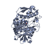
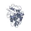
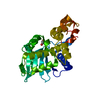
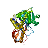
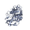
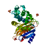
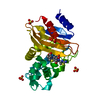
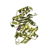
- PDB-2qet: Structure of the mutant S211A of the ribosome inactivating protei... -
+ Open data

- Basic information
| Entry | Database: PDB / ID: 2qet | ||||||
|---|---|---|---|---|---|---|---|
| Title | Structure of the mutant S211A of the ribosome inactivating protein PDL4 from P. dioica in complex with adenine | ||||||
 Components Components | Ribosome-inactivating protein PD-L4 | ||||||
 Keywords Keywords | HYDROLASE / crystal / ribosome inactivating protein | ||||||
| Function / homology |  Function and homology information Function and homology informationrRNA N-glycosylase / rRNA N-glycosylase activity / defense response / toxin activity / negative regulation of translation Similarity search - Function | ||||||
| Biological species |  Phytolacca dioica (plant) Phytolacca dioica (plant) | ||||||
| Method |  X-RAY DIFFRACTION / SYNCHROTRON / MOLECULAR REPLACEMENT / Resolution: 1.24 Å X-RAY DIFFRACTION / SYNCHROTRON / MOLECULAR REPLACEMENT / Resolution: 1.24 Å | ||||||
 Authors Authors | Ruggiero, A. / Berisio, R. | ||||||
 Citation Citation | Journal: Proteins / Year: 2008 Title: Atomic resolution (1.1 A) structure of the ribosome-inactivating protein PD-L4 from Phytolacca dioica L. leaves Authors: Ruggiero, A. / Chambery, A. / Di Maro, A. / Parente, A. / Berisio, R. | ||||||
| History |
|
- Structure visualization
| Structure viewer | Molecule: MolmilJmol/JSmol |
|---|
- Downloads & links
-Download
| PDBx/mmCIF format | 2qet.cif.gz | 137.5 KB | Display | PDBx/mmCIF format |
|---|---|---|---|---|
| PDB format | pdb2qet.ent.gz | 106.7 KB | Display | PDB format |
| PDBx/mmJSON format | 2qet.json.gz | Tree view | PDBx/mmJSON format | |
| Others |  Other downloads Other downloads |
-Validation report
| Arichive directory | https://data.pdbj.org/pub/pdb/validation_reports/qe/2qetftp://data.pdbj.org/pub/pdb/validation_reports/qe/2qet | HTTPS FTP |
|---|
-Related structure data
| Related structure data |  2qesC  2z4uC  2z53C  1qcgS C: citing same article ( S: Starting model for refinement |
|---|---|
| Similar structure data |


-Links
 PDBj
PDBj


- Assembly
| Deposited unit | 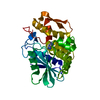
| ||||||||
|---|---|---|---|---|---|---|---|---|---|
| 1 |
| ||||||||
| Unit cell |
|
-Components
| #1: Protein | Mass: 29205.115 Da / Num. of mol.: 1 / Mutation: S211A Source method: isolated from a genetically manipulated source Source: (gene. exp.) Phytolacca dioica (plant) / Plasmid: pET22b(+) / Production host:  |
|---|---|
| #2: Chemical | ChemComp-ADE /   Mass: 135.127 Da / Num. of mol.: 1 / Source method: obtained synthetically / Formula: C5H5N5 Mass: 135.127 Da / Num. of mol.: 1 / Source method: obtained synthetically / Formula: C5H5N5 |
| #3: Water | ChemComp-HOH /  Mass: 18.015 Da / Num. of mol.: 491 / Source method: isolated from a natural source / Formula: H2O Mass: 18.015 Da / Num. of mol.: 491 / Source method: isolated from a natural source / Formula: H2O |
| Has protein modification | Y |
-Experimental details
-Experiment
| Experiment | Method: X-RAY DIFFRACTION / Number of used crystals: 1 |
|---|
- Sample preparation
| Crystal | Density Matthews: 2.1 Å3/Da / Density % sol: 41.56 % |
|---|---|
| Crystal grow | Method: vapor diffusion / Details: VAPOR DIFFUSION |
-Data collection
| Diffraction | Mean temperature: 100 K |
|---|---|
| Diffraction source | Source: SYNCHROTRON / Site: EMBL/DESY, HAMBURG  / Beamline: BW7A / Beamline: BW7A |
| Detector | Type: MAR CCD 165 mm / Detector: CCD / Date: Apr 8, 2006 |
| Radiation | Protocol: SINGLE WAVELENGTH / Monochromatic (M) / Laue (L): M / Scattering type: x-ray |
| Radiation wavelength | Relative weight: 1 |
| Reflection | Resolution: 1.24→30 Å / Num. all: 75869 / Num. obs: 68965 |
| Reflection shell | Highest resolution: 1.24 Å |
- Processing
| Software |
| ||||||||||||||||||||
|---|---|---|---|---|---|---|---|---|---|---|---|---|---|---|---|---|---|---|---|---|---|
| Refinement | Method to determine structure: MOLECULAR REPLACEMENT Starting model: 1qcg Resolution: 1.24→10 Å / Isotropic thermal model: isotropic / Cross valid method: THROUGHOUT / Stereochemistry target values: Engh & Huber
| ||||||||||||||||||||
| Refinement step | Cycle: LAST / Resolution: 1.24→10 Å
| ||||||||||||||||||||
| Refine LS restraints |
|
