 Movie
Movie Controller

[English] 日本語
 Yorodumi
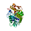
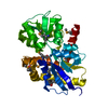
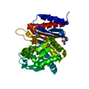
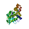
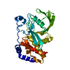
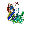
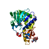
- PDB-1lpc: HIGH RESOLUTION STRUCTURE OF RECOMBINANT DIANTHIN ANTIVIRAL PROTE... -
+ Open data

- Basic information
| Entry | Database: PDB / ID: 1lpc | ||||||
|---|---|---|---|---|---|---|---|
| Title | HIGH RESOLUTION STRUCTURE OF RECOMBINANT DIANTHIN ANTIVIRAL PROTEIN-POTENT ANTI-HIV AGENT (COMPLEX WITH CYCLIC AMP) | ||||||
 Components Components | DIANTHIN 30 | ||||||
 Keywords Keywords | ANTIVIRAL PROTEIN / DIANTHIN ANTIVIRAL PROTEIN / RIBOSOME INACTIVATING PROTEIN / ANTI-HIV AGENT / HIV-1 INTEGRASE INHIBITOR / POLYNUCLEOTIDE:ADENOSINE GLYCOSIDASE | ||||||
| Function / homology |  Function and homology information Function and homology informationregulation of defense response to virus / rRNA N-glycosylase / rRNA N-glycosylase activity / defense response / toxin activity / negative regulation of translation Similarity search - Function | ||||||
| Biological species |  Dianthus caryophyllus (clove pink) Dianthus caryophyllus (clove pink) | ||||||
| Method |  X-RAY DIFFRACTION / MOLECULAR REPLACEMENT / Resolution: 1.7 Å X-RAY DIFFRACTION / MOLECULAR REPLACEMENT / Resolution: 1.7 Å | ||||||
 Authors Authors | Kurinov, I.V. / Rajamohan, F. / Uckun, F.M. | ||||||
 Citation Citation | Journal: Arzneimittelforschung / Year: 2004 Title: High resolution X-ray structure and potent anti-HIV activity of recombinant dianthin antiviral protein. Authors: Kurinov, I.V. / Rajamohan, F. / Uckun, F.M. | ||||||
| History |
|
- Structure visualization
| Structure viewer | Molecule: MolmilJmol/JSmol |
|---|
- Downloads & links
-Download
| PDBx/mmCIF format | 1lpc.cif.gz | 67 KB | Display | PDBx/mmCIF format |
|---|---|---|---|---|
| PDB format | pdb1lpc.ent.gz | 48.1 KB | Display | PDB format |
| PDBx/mmJSON format | 1lpc.json.gz | Tree view | PDBx/mmJSON format | |
| Others |  Other downloads Other downloads |
-Validation report
| Arichive directory | https://data.pdbj.org/pub/pdb/validation_reports/lp/1lpcftp://data.pdbj.org/pub/pdb/validation_reports/lp/1lpc | HTTPS FTP |
|---|
-Related structure data
| Related structure data | 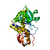 1lp8SC 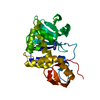 1lpdC S: Starting model for refinement C: citing same article ( |
|---|---|
| Similar structure data |


-Links
 PDBj
PDBj


- Assembly
| Deposited unit | 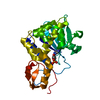
| ||||||||
|---|---|---|---|---|---|---|---|---|---|
| 1 |
| ||||||||
| Unit cell |
|
-Components
| #1: Protein | Mass: 28603.588 Da / Num. of mol.: 1 Source method: isolated from a genetically manipulated source Source: (gene. exp.) Dianthus caryophyllus (clove pink) / Production host:  |
|---|---|
| #2: Chemical | ChemComp-CMP /   Mass: 329.206 Da / Num. of mol.: 1 / Source method: obtained synthetically / Formula: C10H12N5O6P Mass: 329.206 Da / Num. of mol.: 1 / Source method: obtained synthetically / Formula: C10H12N5O6P |
| #3: Water | ChemComp-HOH /  Mass: 18.015 Da / Num. of mol.: 175 / Source method: isolated from a natural source / Formula: H2O Mass: 18.015 Da / Num. of mol.: 175 / Source method: isolated from a natural source / Formula: H2O |
-Experimental details
-Experiment
| Experiment | Method: X-RAY DIFFRACTION / Number of used crystals: 1 |
|---|
- Sample preparation
| Crystal | Density Matthews: 2.1 Å3/Da / Density % sol: 41.41 % |
|---|---|
| Crystal grow | Temperature: 295 K / Method: vapor diffusion, hanging drop / pH: 8 Details: 26% PEG3350, 0.1M CaCl2, pH 8, VAPOR DIFFUSION, HANGING DROP, temperature 295K |
-Data collection
| Diffraction | Mean temperature: 290 K |
|---|---|
| Diffraction source | Source: ROTATING ANODE / Type: RIGAKU RU300 / Wavelength: 1.54 Å |
| Detector | Type: RIGAKU RAXIS IV / Detector: IMAGE PLATE / Date: Dec 23, 2000 |
| Radiation | Monochromator: Yale mirrors / Protocol: SINGLE WAVELENGTH / Monochromatic (M) / Laue (L): M / Scattering type: x-ray |
| Radiation wavelength | Wavelength: 1.54 Å / Relative weight: 1 |
| Reflection | Resolution: 1.7→20 Å / Num. all: 26030 / Num. obs: 24357 / % possible obs: 93.6 % / Observed criterion σ(F): 0 / Observed criterion σ(I): 0 / Rmerge(I) obs: 0.044 / Net I/σ(I): 53.7 |
| Reflection shell | Resolution: 1.7→1.76 Å / Rmerge(I) obs: 0.097 / % possible all: 81.2 |
- Processing
| Software |
| |||||||||||||||||||||||||
|---|---|---|---|---|---|---|---|---|---|---|---|---|---|---|---|---|---|---|---|---|---|---|---|---|---|---|
| Refinement | Method to determine structure: MOLECULAR REPLACEMENT Starting model: PDB ENTRY 1LP8 Resolution: 1.7→8 Å / Isotropic thermal model: Isotropic / Cross valid method: THROUGHOUT / σ(F): 2 / Stereochemistry target values: Engh & Huber
| |||||||||||||||||||||||||
| Displacement parameters | Biso mean: 16.8 Å2 | |||||||||||||||||||||||||
| Refinement step | Cycle: LAST / Resolution: 1.7→8 Å
| |||||||||||||||||||||||||
| Refine LS restraints |
|
