 Movie
Movie Controller


+ Open data

- Basic information
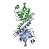
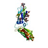
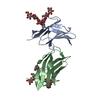
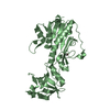
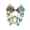
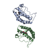
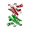
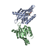
| Entry | Database: PDB / ID: 2qas | ||||||
|---|---|---|---|---|---|---|---|
| Title | Crystal structure of Caulobacter crescentus SspB ortholog | ||||||
 Components Components |
| ||||||
 Keywords Keywords | Hydrolase activator / sspB / adaptor / ClpX / ssrA / UNKNOWN FUNCTION | ||||||
| Function / homology | SspB-like / Stringent starvation protein B / SspB-like superfamily / Stringent starvation protein B / SH3 type barrels. / Roll / Mainly Beta / identical protein binding / Stringent starvation protein B Function and homology information Function and homology information | ||||||
| Biological species |  Caulobacter vibrioides (bacteria) Caulobacter vibrioides (bacteria) | ||||||
| Method |  X-RAY DIFFRACTION / SYNCHROTRON / MOLECULAR REPLACEMENT / Resolution: 2.55 Å X-RAY DIFFRACTION / SYNCHROTRON / MOLECULAR REPLACEMENT / Resolution: 2.55 Å | ||||||
 Authors Authors | Chien, P. / Grant, R.A. / Sauer, R.T. / Baker, T.A. | ||||||
 Citation Citation | Journal: To be Published Title: Crystal structure of Caulobacter crescentus SspB ortholog Authors: Chien, P. / Grant, R.A. / Sauer, R.T. / Baker, T.A. | ||||||
| History |
|
- Structure visualization
| Structure viewer | Molecule: MolmilJmol/JSmol |
|---|
- Downloads & links
-Download
| PDBx/mmCIF format | 2qas.cif.gz | 38.5 KB | Display | PDBx/mmCIF format |
|---|---|---|---|---|
| PDB format | pdb2qas.ent.gz | 26 KB | Display | PDB format |
| PDBx/mmJSON format | 2qas.json.gz | Tree view | PDBx/mmJSON format | |
| Others |  Other downloads Other downloads |
-Validation report
| Arichive directory | https://data.pdbj.org/pub/pdb/validation_reports/qa/2qasftp://data.pdbj.org/pub/pdb/validation_reports/qa/2qas | HTTPS FTP |
|---|
-Related structure data
| Related structure data | |
|---|---|
| Similar structure data |


-Links
 PDBj
PDBj- Assembly
| Deposited unit | 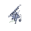
| ||||||||
|---|---|---|---|---|---|---|---|---|---|
| 1 | 
| ||||||||
| Unit cell |
| ||||||||
| Details | Biological assembly is a dimer formed from the monomer in the asymmetric unit by the operations: -x,-y,z + [1,1,0] |
-Components
| #1: Protein | Mass: 17144.199 Da / Num. of mol.: 1 Source method: isolated from a genetically manipulated source Source: (gene. exp.) Caulobacter vibrioides (bacteria) / Strain: CB15N / Gene: CC_2102 / Plasmid: pET28 / Species (production host): Escherichia coli / Production host: |
|---|---|
| #2: Protein/peptide | Mass: 2107.285 Da / Num. of mol.: 1 / Source method: obtained synthetically Details: synthetic ssrA pepitde with N-terminal solubilization tag |
| #3: Water | ChemComp-HOH /  Mass: 18.015 Da / Num. of mol.: 19 / Source method: isolated from a natural source / Formula: H2O Mass: 18.015 Da / Num. of mol.: 19 / Source method: isolated from a natural source / Formula: H2O |
-Experimental details
-Experiment
| Experiment | Method: X-RAY DIFFRACTION / Number of used crystals: 1 |
|---|
- Sample preparation
| Crystal | Density Matthews: 3.07 Å3/Da / Density % sol: 59.99 % |
|---|---|
| Crystal grow | Temperature: 300 K / Method: vapor diffusion, hanging drop / pH: 8 Details: 1.4 M (NH4)2SO4, 0.1 M Tris, pH 8.0, hanging drop, temperature 300K, VAPOR DIFFUSION, HANGING DROP |
-Data collection
| Diffraction | Mean temperature: 110 K |
|---|---|
| Diffraction source | Source: SYNCHROTRON / Site: APS  / Beamline: 24-ID-C / Wavelength: 0.97908 Å / Beamline: 24-ID-C / Wavelength: 0.97908 Å |
| Detector | Type: ADSC QUANTUM 315 / Detector: CCD / Date: Nov 2, 2006 |
| Radiation | Monochromator: crystal monochromator / Protocol: SINGLE WAVELENGTH / Monochromatic (M) / Laue (L): M / Scattering type: x-ray |
| Radiation wavelength | Wavelength: 0.97908 Å / Relative weight: 1 |
| Reflection | Resolution: 2.55→50 Å / Num. all: 8146 / Num. obs: 7817 / % possible obs: 98 % / Observed criterion σ(F): 0 / Observed criterion σ(I): 0 / Redundancy: 10.3 % / Rmerge(I) obs: 0.074 / Χ2: 1.161 / Net I/σ(I): 12.8 |
| Reflection shell | Resolution: 2.55→2.64 Å / Redundancy: 5.8 % / Rmerge(I) obs: 0.377 / Num. unique all: 653 / Χ2: 0.384 / % possible all: 85.2 |
- Processing
| Software |
| ||||||||||||||||||||||||||||||||||||
|---|---|---|---|---|---|---|---|---|---|---|---|---|---|---|---|---|---|---|---|---|---|---|---|---|---|---|---|---|---|---|---|---|---|---|---|---|---|
| Refinement | Method to determine structure: MOLECULAR REPLACEMENT / Resolution: 2.55→26.76 Å / Rfactor Rfree error: 0.01 / Data cutoff high absF: 85886.297 / Data cutoff low absF: 0 / Isotropic thermal model: RESTRAINED / Cross valid method: THROUGHOUT / σ(F): 0 / Stereochemistry target values: Engh & Huber
| ||||||||||||||||||||||||||||||||||||
| Solvent computation | Solvent model: FLAT MODEL / Bsol: 44.361 Å2 / ksol: 0.332 e/Å3 | ||||||||||||||||||||||||||||||||||||
| Displacement parameters | Biso mean: 69.4 Å2
| ||||||||||||||||||||||||||||||||||||
| Refine analyze |
| ||||||||||||||||||||||||||||||||||||
| Refinement step | Cycle: LAST / Resolution: 2.55→26.76 Å
| ||||||||||||||||||||||||||||||||||||
| Refine LS restraints |
| ||||||||||||||||||||||||||||||||||||
| LS refinement shell | Resolution: 2.55→2.71 Å / Rfactor Rfree error: 0.037 / Total num. of bins used: 6
| ||||||||||||||||||||||||||||||||||||
| Xplor file |
|
