 Movie
Movie Controller


+ Open data

- Basic information
| Entry | Database: PDB / ID: 2q9s | ||||||
|---|---|---|---|---|---|---|---|
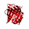
| Title | Linoleic Acid Bound to Fatty Acid Binding Protein 4 | ||||||
 Components Components | Fatty acid-binding protein | ||||||
 Keywords Keywords | LIPID BINDING PROTEIN / beta clamshell | ||||||
| Function / homology |  Function and homology information Function and homology informationTriglyceride catabolism / hormone receptor binding / long-chain fatty acid transmembrane transporter activity / long-chain fatty acid binding / cellular response to lithium ion / white fat cell differentiation / long-chain fatty acid transport / brown fat cell differentiation / cholesterol homeostasis / response to bacterium ...Triglyceride catabolism / hormone receptor binding / long-chain fatty acid transmembrane transporter activity / long-chain fatty acid binding / cellular response to lithium ion / white fat cell differentiation / long-chain fatty acid transport / brown fat cell differentiation / cholesterol homeostasis / response to bacterium / positive regulation of inflammatory response / cellular response to tumor necrosis factor / positive regulation of cold-induced thermogenesis / negative regulation of DNA-templated transcription / nucleoplasm / nucleus / cytoplasm Similarity search - Function | ||||||
| Biological species |  | ||||||
| Method |  X-RAY DIFFRACTION / SYNCHROTRON / MOLECULAR REPLACEMENT / Resolution: 2.3 Å X-RAY DIFFRACTION / SYNCHROTRON / MOLECULAR REPLACEMENT / Resolution: 2.3 Å | ||||||
 Authors Authors | Gillilan, R.E. / Ayers, S.D. / Noy, N. | ||||||
 Citation Citation | Journal: J.Mol.Biol. / Year: 2007 Title: Structural Basis for Activation of Fatty Acid-binding Protein 4. Authors: Gillilan, R.E. / Ayers, S.D. / Noy, N. | ||||||
| History |
|
- Structure visualization
| Structure viewer | Molecule: MolmilJmol/JSmol |
|---|
- Downloads & links
-Download
| PDBx/mmCIF format | 2q9s.cif.gz | 41.4 KB | Display | PDBx/mmCIF format |
|---|---|---|---|---|
| PDB format | pdb2q9s.ent.gz | 27.9 KB | Display | PDB format |
| PDBx/mmJSON format | 2q9s.json.gz | Tree view | PDBx/mmJSON format | |
| Others |  Other downloads Other downloads |
-Validation report
| Summary document | 2q9s_validation.pdf.gz | 798.1 KB | Display | wwPDB validaton report |
|---|---|---|---|---|
| Full document | 2q9s_full_validation.pdf.gz | 799.1 KB | Display | |
| Data in XML | 2q9s_validation.xml.gz | 8.1 KB | Display | |
| Data in CIF | 2q9s_validation.cif.gz | 10.1 KB | Display | |
| Arichive directory | https://data.pdbj.org/pub/pdb/validation_reports/q9/2q9sftp://data.pdbj.org/pub/pdb/validation_reports/q9/2q9s | HTTPS FTP |
-Related structure data


-Links
 PDBj
PDBj

- Assembly
| Deposited unit | 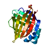
| ||||||||
|---|---|---|---|---|---|---|---|---|---|
| 1 | 
| ||||||||
| 2 | 
| ||||||||
| Unit cell |
| ||||||||
| Components on special symmetry positions |
| ||||||||
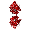
| Details | The assembly is a dimer. The second half is generated by [-x,y,1/2-z] |
-Components
| #1: Protein | Mass: 17001.518 Da / Num. of mol.: 1 Source method: isolated from a genetically manipulated source Source: (gene. exp.)  |
|---|---|
| #2: Chemical | ChemComp-SO4 /   Mass: 96.063 Da / Num. of mol.: 1 / Source method: obtained synthetically / Formula: SO4 Mass: 96.063 Da / Num. of mol.: 1 / Source method: obtained synthetically / Formula: SO4 |
| #3: Chemical | ChemComp-EIC /   Mass: 280.445 Da / Num. of mol.: 1 / Source method: obtained synthetically / Formula: C18H32O2 Mass: 280.445 Da / Num. of mol.: 1 / Source method: obtained synthetically / Formula: C18H32O2 |
| #4: Water | ChemComp-HOH /  Mass: 18.015 Da / Num. of mol.: 48 / Source method: isolated from a natural source / Formula: H2O Mass: 18.015 Da / Num. of mol.: 48 / Source method: isolated from a natural source / Formula: H2O |
-Experimental details
-Experiment
| Experiment | Method: X-RAY DIFFRACTION / Number of used crystals: 1 |
|---|
- Sample preparation
| Crystal | Density Matthews: 2.74 Å3/Da / Density % sol: 55.09 % |
|---|---|
| Crystal grow | Temperature: 293 K / Method: vapor diffusion, hanging drop / pH: 6.5 Details: 0.1 M Sodium Cacodylate, 0.1 M Ammonium Sulfate, 35% PEG 8000, pH 6.5, VAPOR DIFFUSION, HANGING DROP, temperature 293K |
-Data collection
| Diffraction | Mean temperature: 100 K |
|---|---|
| Diffraction source | Source: SYNCHROTRON / Site: CHESS  / Beamline: F1 / Wavelength: 0.9176 Å / Beamline: F1 / Wavelength: 0.9176 Å |
| Detector | Type: ADSC QUANTUM 4 / Detector: CCD / Date: Apr 30, 2006 / Details: mirrors |
| Radiation | Protocol: SINGLE WAVELENGTH / Monochromatic (M) / Laue (L): M / Scattering type: x-ray |
| Radiation wavelength | Wavelength: 0.9176 Å / Relative weight: 1 |
| Reflection | Resolution: 2.3→50 Å / Num. obs: 8446 / % possible obs: 98.7 % / Redundancy: 9.4 % / Rmerge(I) obs: 0.082 / Χ2: 1.166 / Net I/σ(I): 24.7 |
| Reflection shell | Resolution: 2.3→2.38 Å / Redundancy: 9.6 % / Rmerge(I) obs: 0.176 / Num. unique all: 834 / Χ2: 1.158 / % possible all: 99.3 |
- Processing
| Software |
| ||||||||||||||||||||||||||||||||||||||||||||||||||||||||||||||||||||||||||||||||||||||||||||||||||||
|---|---|---|---|---|---|---|---|---|---|---|---|---|---|---|---|---|---|---|---|---|---|---|---|---|---|---|---|---|---|---|---|---|---|---|---|---|---|---|---|---|---|---|---|---|---|---|---|---|---|---|---|---|---|---|---|---|---|---|---|---|---|---|---|---|---|---|---|---|---|---|---|---|---|---|---|---|---|---|---|---|---|---|---|---|---|---|---|---|---|---|---|---|---|---|---|---|---|---|---|---|---|
| Refinement | Method to determine structure: MOLECULAR REPLACEMENT / Resolution: 2.3→12 Å / Cor.coef. Fo:Fc: 0.937 / Cor.coef. Fo:Fc free: 0.906 / SU B: 6.819 / SU ML: 0.165 / TLS residual ADP flag: LIKELY RESIDUAL / Cross valid method: THROUGHOUT / σ(F): 0 / ESU R: 0.287 / ESU R Free: 0.228 / Stereochemistry target values: MAXIMUM LIKELIHOOD / Details: HYDROGENS HAVE BEEN ADDED IN THE RIDING POSITIONS
| ||||||||||||||||||||||||||||||||||||||||||||||||||||||||||||||||||||||||||||||||||||||||||||||||||||
| Solvent computation | Ion probe radii: 0.8 Å / Shrinkage radii: 0.8 Å / VDW probe radii: 1.4 Å / Solvent model: BABINET MODEL WITH MASK | ||||||||||||||||||||||||||||||||||||||||||||||||||||||||||||||||||||||||||||||||||||||||||||||||||||
| Displacement parameters | Biso mean: 25.784 Å2
| ||||||||||||||||||||||||||||||||||||||||||||||||||||||||||||||||||||||||||||||||||||||||||||||||||||
| Refinement step | Cycle: LAST / Resolution: 2.3→12 Å
| ||||||||||||||||||||||||||||||||||||||||||||||||||||||||||||||||||||||||||||||||||||||||||||||||||||
| Refine LS restraints |
| ||||||||||||||||||||||||||||||||||||||||||||||||||||||||||||||||||||||||||||||||||||||||||||||||||||
| LS refinement shell | Resolution: 2.3→2.42 Å / Total num. of bins used: 10
| ||||||||||||||||||||||||||||||||||||||||||||||||||||||||||||||||||||||||||||||||||||||||||||||||||||
| Refinement TLS params. | Method: refined / Refine-ID: X-RAY DIFFRACTION
| ||||||||||||||||||||||||||||||||||||||||||||||||||||||||||||||||||||||||||||||||||||||||||||||||||||
| Refinement TLS group |
|
