 Movie
Movie Controller

[English] 日本語
 Yorodumi
- PDB-2q3c: 2.1 A Resolution Crystal Structure of O-Acetylserine Sulfhydrylas... -
+ Open data

- Basic information
| Entry | Database: PDB / ID: 2q3c | ||||||
|---|---|---|---|---|---|---|---|
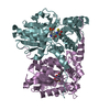
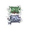
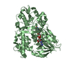
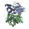
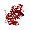
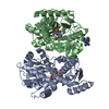
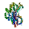
| Title | 2.1 A Resolution Crystal Structure of O-Acetylserine Sulfhydrylase (OASS) Holoenzyme From MYCOBACTERIUM TUBERCULOSIS in Complex with the Inhibitory Peptide DFSI | ||||||
 Components Components |
| ||||||
 Keywords Keywords | TRANSFERASE / Mycobacterium tuberculosis / Pyridoxal-5'-phosphate / sulphur metabolism / cysteine biosynthesis / SAT / peptide-inhibitor | ||||||
| Function / homology |  Function and homology information Function and homology informationCysteine synthesis from O-acetylserine / L-cysteine desulfhydrase activity / cysteine synthase / cysteine synthase activity / L-cysteine biosynthetic process / : / pyridoxal phosphate binding / extracellular region / cytosol / cytoplasm Similarity search - Function | ||||||
| Biological species |   Mycobacterium tuberculosis (bacteria) Mycobacterium tuberculosis (bacteria) | ||||||
| Method |  X-RAY DIFFRACTION / SYNCHROTRON / MOLECULAR REPLACEMENT / Resolution: 2.1 Å X-RAY DIFFRACTION / SYNCHROTRON / MOLECULAR REPLACEMENT / Resolution: 2.1 Å | ||||||
 Authors Authors | Schneider, G. / Schnell, R. | ||||||
 Citation Citation | Journal: J.Biol.Chem. / Year: 2007 Title: Structural Insights into Catalysis and Inhibition of O-Acetylserine Sulfhydrylase from Mycobacterium tuberculosis: CRYSTAL STRUCTURES OF THE ENZYME {alpha}-AMINOACRYLATE INTERMEDIATE AND AN ...Title: Structural Insights into Catalysis and Inhibition of O-Acetylserine Sulfhydrylase from Mycobacterium tuberculosis: CRYSTAL STRUCTURES OF THE ENZYME {alpha}-AMINOACRYLATE INTERMEDIATE AND AN ENZYME-INHIBITOR COMPLEX. Authors: Schnell, R. / Oehlmann, W. / Singh, M. / Schneider, G. | ||||||
| History |
|
- Structure visualization
| Structure viewer | Molecule: MolmilJmol/JSmol |
|---|
- Downloads & links
-Download
| PDBx/mmCIF format | 2q3c.cif.gz | 76.4 KB | Display | PDBx/mmCIF format |
|---|---|---|---|---|
| PDB format | pdb2q3c.ent.gz | 54.6 KB | Display | PDB format |
| PDBx/mmJSON format | 2q3c.json.gz | Tree view | PDBx/mmJSON format | |
| Others |  Other downloads Other downloads |
-Validation report
| Arichive directory | https://data.pdbj.org/pub/pdb/validation_reports/q3/2q3cftp://data.pdbj.org/pub/pdb/validation_reports/q3/2q3c | HTTPS FTP |
|---|
-Related structure data
| Related structure data |  2q3bSC 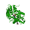 2q3dC S: Starting model for refinement C: citing same article ( |
|---|---|
| Similar structure data |


-Links
 PDBj
PDBj
- Assembly
| Deposited unit | 
| ||||||||
|---|---|---|---|---|---|---|---|---|---|
| 1 | 
| ||||||||
| Unit cell |
| ||||||||
| Details | The OASS forms a dimer, the second part of the biological assembly is generated by the two fold axis: -y, x, -z. |
-Components
| #1: Protein | Mass: 33296.008 Da / Num. of mol.: 1 Source method: isolated from a genetically manipulated source Source: (gene. exp.) Mycobacterium tuberculosis (bacteria) / Strain: Rv2334 / Gene: cysK / Plasmid: pET28a / Species (production host): Escherichia coli / Production host: References: UniProt: P0A534, UniProt: P9WP55*PLUS, cysteine synthase |
|---|---|
| #2: Protein/peptide | Mass: 480.512 Da / Num. of mol.: 1 / Source method: obtained synthetically / Source: (synth.) Mycobacterium tuberculosis (bacteria) |
| #3: Chemical | ChemComp-MPD / (  Mass: 118.174 Da / Num. of mol.: 1 / Source method: obtained synthetically / Formula: C6H14O2 / Comment: precipitant*YM Mass: 118.174 Da / Num. of mol.: 1 / Source method: obtained synthetically / Formula: C6H14O2 / Comment: precipitant*YM |
| #4: Water | ChemComp-HOH /  Mass: 18.015 Da / Num. of mol.: 163 / Source method: isolated from a natural source / Formula: H2O Mass: 18.015 Da / Num. of mol.: 163 / Source method: isolated from a natural source / Formula: H2O |
-Experimental details
-Experiment
| Experiment | Method: X-RAY DIFFRACTION / Number of used crystals: 1 |
|---|
- Sample preparation
| Crystal | Density Matthews: 3.61 Å3/Da / Density % sol: 65.96 % |
|---|---|
| Crystal grow | Temperature: 293 K / Method: vapor diffusion, hanging drop / pH: 8 Details: 0.1 M HEPES, 80% MPD, 4 mM DFSI-peptide, pH 8.0, VAPOR DIFFUSION, HANGING DROP, temperature 293K |
-Data collection
| Diffraction | Mean temperature: 110 K |
|---|---|
| Diffraction source | Source: SYNCHROTRON / Site: ESRF  / Beamline: ID14-1 / Wavelength: 0.934 Å / Beamline: ID14-1 / Wavelength: 0.934 Å |
| Detector | Type: ADSC QUANTUM 210 / Detector: CCD / Date: Mar 3, 2007 |
| Radiation | Monochromator: Diamond(111), Ge(220) / Protocol: SINGLE WAVELENGTH / Monochromatic (M) / Laue (L): M / Scattering type: x-ray |
| Radiation wavelength | Wavelength: 0.934 Å / Relative weight: 1 |
| Reflection | Resolution: 2.1→59.66 Å / Num. all: 26571 / Num. obs: 26571 / % possible obs: 93.2 % / Observed criterion σ(F): 0 / Observed criterion σ(I): 0 / Redundancy: 5.1 % / Biso Wilson estimate: 31 Å2 / Rmerge(I) obs: 0.089 / Net I/σ(I): 11.5 |
| Reflection shell | Resolution: 2.1→2.21 Å / Redundancy: 5 % / Rmerge(I) obs: 0.352 / Mean I/σ(I) obs: 3.3 / Num. unique all: 3941 / % possible all: 96.2 |
- Processing
| Software |
| ||||||||||||||||||||||||||||||||||||||||||||||||||||||||||||||||||||||||||||||||||||||||||||||||||||||||||||||
|---|---|---|---|---|---|---|---|---|---|---|---|---|---|---|---|---|---|---|---|---|---|---|---|---|---|---|---|---|---|---|---|---|---|---|---|---|---|---|---|---|---|---|---|---|---|---|---|---|---|---|---|---|---|---|---|---|---|---|---|---|---|---|---|---|---|---|---|---|---|---|---|---|---|---|---|---|---|---|---|---|---|---|---|---|---|---|---|---|---|---|---|---|---|---|---|---|---|---|---|---|---|---|---|---|---|---|---|---|---|---|---|
| Refinement | Method to determine structure: MOLECULAR REPLACEMENT Starting model: pdb entry 2Q3B, Holoenzyme Resolution: 2.1→51.23 Å / Cor.coef. Fo:Fc: 0.959 / Cor.coef. Fo:Fc free: 0.944 / SU B: 4.219 / SU ML: 0.108 / Cross valid method: THROUGHOUT / σ(F): 0 / σ(I): 0 / ESU R: 0.165 / ESU R Free: 0.142 / Stereochemistry target values: MAXIMUM LIKELIHOOD / Details: HYDROGENS HAVE BEEN ADDED IN THE RIDING POSITIONS
| ||||||||||||||||||||||||||||||||||||||||||||||||||||||||||||||||||||||||||||||||||||||||||||||||||||||||||||||
| Solvent computation | Ion probe radii: 0.8 Å / Shrinkage radii: 0.8 Å / VDW probe radii: 1.2 Å / Solvent model: BABINET MODEL WITH MASK | ||||||||||||||||||||||||||||||||||||||||||||||||||||||||||||||||||||||||||||||||||||||||||||||||||||||||||||||
| Displacement parameters | Biso mean: 36.734 Å2
| ||||||||||||||||||||||||||||||||||||||||||||||||||||||||||||||||||||||||||||||||||||||||||||||||||||||||||||||
| Refinement step | Cycle: LAST / Resolution: 2.1→51.23 Å
| ||||||||||||||||||||||||||||||||||||||||||||||||||||||||||||||||||||||||||||||||||||||||||||||||||||||||||||||
| Refine LS restraints |
| ||||||||||||||||||||||||||||||||||||||||||||||||||||||||||||||||||||||||||||||||||||||||||||||||||||||||||||||
| LS refinement shell | Resolution: 2.1→2.155 Å / Total num. of bins used: 20
|
