 Movie
Movie Controller


+ Open data

- Basic information
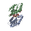
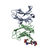
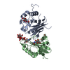
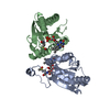
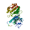
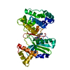
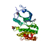
| Entry | Database: PDB / ID: 2q2k | ||||||
|---|---|---|---|---|---|---|---|
| Title | Structure of nucleic-acid binding protein | ||||||
 Components Components |
| ||||||
 Keywords Keywords | DNA BINDING PROTEIN/DNA / protein-DNA / partition / segregation / parB / DNA BINDING PROTEIN-DNA COMPLEX | ||||||
| Function / homology | Arc Repressor Mutant - #20 / Arc Repressor Mutant / Helix non-globular / Special / DNA binding / DNA / DNA (> 10) / Conserved domain protein / Uncharacterized protein Function and homology information Function and homology information | ||||||
| Biological species |   Staphylococcus aureus (bacteria) Staphylococcus aureus (bacteria) | ||||||
| Method |  X-RAY DIFFRACTION / SYNCHROTRON / MIR / Resolution: 3 Å X-RAY DIFFRACTION / SYNCHROTRON / MIR / Resolution: 3 Å | ||||||
 Authors Authors | Schumacher, M.A. / Glover, T. / Firth, N. | ||||||
 Citation Citation | Journal: Nature / Year: 2007 Title: Segrosome structure revealed by a complex of ParR with centromere DNA. Authors: Schumacher, M.A. / Glover, T.C. / Brzoska, A.J. / Jensen, S.O. / Dunham, T.D. / Skurray, R.A. / Firth, N. | ||||||
| History |
|
- Structure visualization
| Structure viewer | Molecule: MolmilJmol/JSmol |
|---|
- Downloads & links
-Download
| PDBx/mmCIF format | 2q2k.cif.gz | 46.6 KB | Display | PDBx/mmCIF format |
|---|---|---|---|---|
| PDB format | pdb2q2k.ent.gz | 30.1 KB | Display | PDB format |
| PDBx/mmJSON format | 2q2k.json.gz | Tree view | PDBx/mmJSON format | |
| Others |  Other downloads Other downloads |
-Validation report
| Arichive directory | https://data.pdbj.org/pub/pdb/validation_reports/q2/2q2kftp://data.pdbj.org/pub/pdb/validation_reports/q2/2q2k | HTTPS FTP |
|---|
-Related structure data
| Similar structure data |
|---|


-Links
 PDBj
PDBj


- Assembly
| Deposited unit | 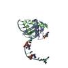
| ||||||||
|---|---|---|---|---|---|---|---|---|---|
| 1 | 
| ||||||||
| Unit cell |
|
-Components
| #1: DNA chain | Mass: 6354.755 Da / Num. of mol.: 1 / Source method: obtained synthetically | ||
|---|---|---|---|
| #2: Protein | Mass: 8167.265 Da / Num. of mol.: 2 Source method: isolated from a genetically manipulated source Source: (gene. exp.) Staphylococcus aureus (bacteria) / Plasmid: pET15b / Species (production host): Escherichia coli / Production host: #3: Chemical | ChemComp-EPE / |   Mass: 238.305 Da / Num. of mol.: 1 / Source method: obtained synthetically / Formula: C8H18N2O4S / Comment: pH buffer*YM Mass: 238.305 Da / Num. of mol.: 1 / Source method: obtained synthetically / Formula: C8H18N2O4S / Comment: pH buffer*YM |
-Experimental details
-Experiment
| Experiment | Method: X-RAY DIFFRACTION / Number of used crystals: 1 |
|---|
- Sample preparation
| Crystal | Density Matthews: 2.32 Å3/Da / Density % sol: 47.06 % | ||||||||||||||||||||||||||||
|---|---|---|---|---|---|---|---|---|---|---|---|---|---|---|---|---|---|---|---|---|---|---|---|---|---|---|---|---|---|
| Crystal grow | Temperature: 298 K / Method: vapor diffusion, hanging drop / pH: 7.5 Details: PEG 4000, isopropanol, HEPES, pH 7.5, VAPOR DIFFUSION, HANGING DROP, temperature 298K | ||||||||||||||||||||||||||||
| Components of the solutions |
|
-Data collection
| Diffraction | Mean temperature: 100 K |
|---|---|
| Diffraction source | Source: SYNCHROTRON / Site: ALS  / Beamline: 8.3.1 / Wavelength: 1.03 Å / Beamline: 8.3.1 / Wavelength: 1.03 Å |
| Detector | Type: ADSC QUANTUM 4 / Detector: CCD / Date: Apr 4, 2007 / Details: mirrors |
| Radiation | Monochromator: graphite / Protocol: SINGLE WAVELENGTH / Monochromatic (M) / Laue (L): M / Scattering type: x-ray |
| Radiation wavelength | Wavelength: 1.03 Å / Relative weight: 1 |
| Reflection | Resolution: 3→77.6 Å / Num. all: 4650 / Num. obs: 4506 / % possible obs: 97 % / Observed criterion σ(F): 0 / Observed criterion σ(I): 0 / Redundancy: 3 % / Biso Wilson estimate: 87 Å2 / Rmerge(I) obs: 0.058 / Rsym value: 0.06 / Net I/σ(I): 15 |
| Reflection shell | Resolution: 3→3.19 Å / Redundancy: 3 % / Rmerge(I) obs: 0.372 / Mean I/σ(I) obs: 2.1 / Num. unique all: 450 / Rsym value: 0.365 / % possible all: 97 |
- Processing
| Software |
| ||||||||||||||||||||||||||||||||||||
|---|---|---|---|---|---|---|---|---|---|---|---|---|---|---|---|---|---|---|---|---|---|---|---|---|---|---|---|---|---|---|---|---|---|---|---|---|---|
| Refinement | Method to determine structure: MIR / Resolution: 3→41.27 Å / Rfactor Rfree error: 0.014 / Data cutoff high absF: 1236191.32 / Data cutoff low absF: 0 / Isotropic thermal model: RESTRAINED / Cross valid method: THROUGHOUT / σ(F): 0 / σ(I): 0 / Stereochemistry target values: Engh & Huber
| ||||||||||||||||||||||||||||||||||||
| Solvent computation | Solvent model: FLAT MODEL / Bsol: 50.0649 Å2 / ksol: 0.342795 e/Å3 | ||||||||||||||||||||||||||||||||||||
| Displacement parameters | Biso mean: 77.4 Å2
| ||||||||||||||||||||||||||||||||||||
| Refine analyze |
| ||||||||||||||||||||||||||||||||||||
| Refinement step | Cycle: LAST / Resolution: 3→41.27 Å
| ||||||||||||||||||||||||||||||||||||
| Refine LS restraints |
| ||||||||||||||||||||||||||||||||||||
| LS refinement shell | Resolution: 3→3.19 Å / Rfactor Rfree error: 0.061 / Total num. of bins used: 6
| ||||||||||||||||||||||||||||||||||||
| Xplor file |
|
