regulation of nitric oxide metabolic process / Derlin-1-VIMP complex / negative regulation of acute inflammatory response to antigenic stimulus / negative regulation of macrophage apoptotic process / negative regulation of glycogen biosynthetic process / negative regulation of D-glucose import across plasma membrane / low-density lipoprotein particle / very-low-density lipoprotein particle / Derlin-1 retrotranslocation complex / response to redox state ...regulation of nitric oxide metabolic process / Derlin-1-VIMP complex / negative regulation of acute inflammatory response to antigenic stimulus / negative regulation of macrophage apoptotic process / negative regulation of glycogen biosynthetic process / negative regulation of D-glucose import across plasma membrane / low-density lipoprotein particle / very-low-density lipoprotein particle / Derlin-1 retrotranslocation complex / response to redox state / regulation of gluconeogenesis / retrograde protein transport, ER to cytosol / ER overload response / antioxidant activity / ubiquitin-specific protease binding / negative regulation of interleukin-6 production / negative regulation of tumor necrosis factor production / response to glucose / negative regulation of endoplasmic reticulum stress-induced intrinsic apoptotic signaling pathway / cytoplasmic microtubule / endoplasmic reticulum unfolded protein response / ERAD pathway / cell redox homeostasis / establishment of protein localization / negative regulation of inflammatory response / cellular response to insulin stimulus / E3 ubiquitin ligases ubiquitinate target proteins / cellular response to lipopolysaccharide / ATPase binding / signaling receptor activity / cellular response to oxidative stress / endoplasmic reticulum membrane / enzyme binding / endoplasmic reticulum / plasma membrane Similarity search - Function
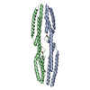
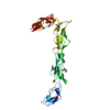
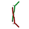
Single alpha-helices involved in coiled-coils or other helix-helix interfaces - #2950 / Selenoprotein S / Selenoprotein S (SelS) / Single alpha-helices involved in coiled-coils or other helix-helix interfaces / Helix non-globular / Special Similarity search - Domain/homology
Resolution: 1.5→1.55 Å / Redundancy: 5.8 % / Mean I/σ(I) obs: 4.2 / Num. unique all: 1216 / Rsym value: 0.325 / % possible all: 88
-
Processing
Software
Name
Version
Classification
REFMAC
5.2.0019
refinement
HKL-2000
datacollection
HKL-2000
datareduction
HKL-2000
datascaling
SOLVE
phasing
Refinement
Method to determine structure: SAD / Resolution: 1.5→31.2 Å / Cor.coef. Fo:Fc: 0.963 / Cor.coef. Fo:Fc free: 0.95 / SU B: 2.658 / SU ML: 0.05 / Cross valid method: THROUGHOUT / ESU R: 0.074 / ESU R Free: 0.077 / Stereochemistry target values: MAXIMUM LIKELIHOOD Details: HYDROGENS HAVE BEEN ADDED IN THE RIDING POSITIONS. ATOM RECORD CONTAINS SUM OF TLS AND RESIDUAL B FACTORS. ANISOU RECORD CONTAINS SUM OF TLS AND RESIDUAL U FACTORS.
Rfactor
Num. reflection
% reflection
Selection details
Rfree
0.21434
668
4.9 %
RANDOM
Rwork
0.18135
-
-
-
all
0.18291
-
-
-
obs
0.18291
12947
97.9 %
-
Solvent computation
Ion probe radii: 0.8 Å / Shrinkage radii: 0.8 Å / VDW probe radii: 1.4 Å / Solvent model: BABINET MODEL WITH MASK
Displacement parameters
Biso mean: 21.637 Å2
Baniso -1
Baniso -2
Baniso -3
1-
0.43 Å2
0 Å2
2.12 Å2
2-
-
-0.46 Å2
0 Å2
3-
-
-
0.29 Å2
Refinement step
Cycle: LAST / Resolution: 1.5→31.2 Å
Protein
Nucleic acid
Ligand
Solvent
Total
Num. atoms
577
0
8
68
653
Refine LS restraints
Refine-ID
Type
Dev ideal
Dev ideal target
Number
X-RAY DIFFRACTION
r_bond_refined_d
0.016
0.022
641
X-RAY DIFFRACTION
r_angle_refined_deg
1.498
1.984
858
X-RAY DIFFRACTION
r_dihedral_angle_1_deg
3.675
5
82
X-RAY DIFFRACTION
r_dihedral_angle_2_deg
30.99
22.667
30
X-RAY DIFFRACTION
r_dihedral_angle_3_deg
14.956
15
142
X-RAY DIFFRACTION
r_dihedral_angle_4_deg
15.702
15
10
X-RAY DIFFRACTION
r_chiral_restr
0.099
0.2
92
X-RAY DIFFRACTION
r_gen_planes_refined
0.007
0.02
478
X-RAY DIFFRACTION
r_nbd_refined
0.217
0.2
292
X-RAY DIFFRACTION
r_nbtor_refined
0.286
0.2
464
X-RAY DIFFRACTION
r_xyhbond_nbd_refined
0.121
0.2
37
X-RAY DIFFRACTION
r_symmetry_vdw_refined
0.244
0.2
75
X-RAY DIFFRACTION
r_symmetry_hbond_refined
0.189
0.2
19
X-RAY DIFFRACTION
r_mcbond_it
1.856
3
411
X-RAY DIFFRACTION
r_mcangle_it
2.356
4
636
X-RAY DIFFRACTION
r_scbond_it
4.771
5
255
X-RAY DIFFRACTION
r_scangle_it
6.453
7
222
LS refinement shell
Resolution: 1.5→1.54 Å / Total num. of bins used: 20
Rfactor
Num. reflection
% reflection
Rfree
0.183
39
-
Rwork
0.18
816
-
obs
-
-
84.4 %
Refinement TLS params.
Method: refined / Refine-ID: X-RAY DIFFRACTION
ID
L11 (°2)
L12 (°2)
L13 (°2)
L22 (°2)
L23 (°2)
L33 (°2)
S11 (Å °)
S12 (Å °)
S13 (Å °)
S21 (Å °)
S22 (Å °)
S23 (Å °)
S31 (Å °)
S32 (Å °)
S33 (Å °)
T11 (Å2)
T12 (Å2)
T13 (Å2)
T22 (Å2)
T23 (Å2)
T33 (Å2)
Origin x (Å)
Origin y (Å)
Origin z (Å)
1
5.9482
1.1038
2.0383
0.6346
0.716
5.2116
-0.1732
0.1692
0.2357
0.2202
0.0005
-0.1566
-0.47
0.3229
0.1727
0.1136
-0.0488
-0.0309
0.0942
0.059
0.0831
35.5783
9.3324
15.0445
2
0.0757
0.8181
-0.7316
9.0769
-8.711
9.8449
-0.0884
-0.0236
0.0059
0.0342
-0.2071
-0.0627
0.02
0.4013
0.2955
0.113
-0.0193
-0.0448
0.1068
0.0273
0.0523
33.1033
2.6669
19.5414
3
2.6643
0.5103
-1.7005
1.2835
-1.9206
8.5385
0.1086
-0.0258
0.0572
0.0177
-0.1475
0.0037
-0.1472
0.0106
0.0389
0.1202
0.004
-0.0331
0.073
0.0111
0.0621
30.7237
-1.4304
25.2265
4
3.605
1.1063
-5.4263
3.3224
-3.3597
9.1303
-0.2602
-0.1133
-0.1661
-0.3228
0.1109
-0.1144
0.5219
-0.0552
0.1493
0.1136
-0.0114
-0.0123
0.0774
0.0069
0.0631
30.5435
-9.0851
33.4654
5
1.2439
2.6621
-0.2772
5.9938
-1.0222
0.6827
-0.2528
0.0418
-0.005
-0.0292
0.2182
0.0047
0.0612
-0.0074
0.0346
0.0675
-0.006
-0.0279
0.0911
0.0009
0.0917
25.2222
-8.9463
39.8312
6
5.5306
-0.4778
-3.2422
1.1259
-0.693
3.0968
0.046
0.1132
0.0196
-0.0232
-0.093
0.1513
-0.1344
-0.1762
0.047
0.0711
-0.0008
0.0027
0.0806
-0.0139
0.1075
18.2802
-11.5003
45.2245
7
24.1225
-1.8224
-11.3643
11.5571
1.2021
5.8614
0.2598
-0.3071
0.4205
-0.1556
-0.2063
0.0481
-0.2009
-0.5047
-0.0534
0.0118
0.0163
0.0168
0.1269
-0.0346
0.0754
12.738
-10.6834
51.0636
8
30.6602
5.0544
-10.5725
2.5851
-4.2348
7.1904
-0.4106
-0.6096
-0.5909
-0.0025
0.0125
0.0457
0.1927
0.1778
0.398
0.024
0.0257
0.043
0.1532
-0.0024
0.0933
6.0279
-14.207
55.446
9
26.6841
0.3141
-10.0274
2.0588
1.6266
6.1599
0.0069
-0.5938
0.0421
-0.1193
0.0306
0.0601
-0.4376
0.2225
-0.0375
-0.0815
0.0266
0.1275
0.1729
0.0342
0.0341
-1.3768
-14.4323
60.3777
10
20.3293
3.2243
-12.1708
12.1557
-10.9418
30.7241
-0.0129
-1.2932
-0.8402
0.6382
-0.3629
-0.5052
0.5945
1.154
0.3758
0.0828
0.0307
-0.0021
0.24
0.0408
0.1419
-7.2911
-17.101
66.4458
11
29.0737
13.3046
-20.3258
8.4209
-11.7453
23.5304
0.5071
-1.2466
0.657
0.1547
-0.646
0.3888
-0.1511
1.5634
0.1389
-0.0359
-0.0411
0.0658
0.2093
-0.0061
0.08
-15.4327
-17.3557
71.4811
12
31.195
3.6741
-11.5087
11.2599
-14.6479
20.5648
1.4451
-1.6585
0.7222
-0.2521
-0.0705
0.8866
-0.8359
0.2641
-1.3746
0.0159
-0.0652
0.0777
0.1195
0.0273
0.1406
-23.7756
-19.6142
77.0238
Refinement TLS group
ID
Refine-ID
Refine TLS-ID
Auth asym-ID
Label asym-ID
Auth seq-ID
Label seq-ID
1
X-RAY DIFFRACTION
1
A
A
51 - 58
18 - 25
2
X-RAY DIFFRACTION
2
A
A
59 - 62
26 - 29
3
X-RAY DIFFRACTION
3
A
A
63 - 69
30 - 36
4
X-RAY DIFFRACTION
4
A
A
70 - 76
37 - 43
5
X-RAY DIFFRACTION
5
A
A
77 - 81
44 - 48
6
X-RAY DIFFRACTION
6
A
A
82 - 87
49 - 54
7
X-RAY DIFFRACTION
7
A
A
88 - 92
55 - 59
8
X-RAY DIFFRACTION
8
A
A
93 - 98
60 - 65
9
X-RAY DIFFRACTION
9
A
A
99 - 104
66 - 71
10
X-RAY DIFFRACTION
10
A
A
105 - 109
72 - 76
11
X-RAY DIFFRACTION
11
A
A
110 - 117
77 - 84
12
X-RAY DIFFRACTION
12
A
A
118 - 122
85 - 89
+
About Yorodumi
-
News
-
Feb 9, 2022. New format data for meta-information of EMDB entries
New format data for meta-information of EMDB entries
Version 3 of the EMDB header file is now the official format.
The previous official version 1.9 will be removed from the archive.
In the structure databanks used in Yorodumi, some data are registered as the other names, "COVID-19 virus" and "2019-nCoV". Here are the details of the virus and the list of structure data.
Jan 31, 2019. EMDB accession codes are about to change! (news from PDBe EMDB page)
EMDB accession codes are about to change! (news from PDBe EMDB page)
The allocation of 4 digits for EMDB accession codes will soon come to an end. Whilst these codes will remain in use, new EMDB accession codes will include an additional digit and will expand incrementally as the available range of codes is exhausted. The current 4-digit format prefixed with “EMD-” (i.e. EMD-XXXX) will advance to a 5-digit format (i.e. EMD-XXXXX), and so on. It is currently estimated that the 4-digit codes will be depleted around Spring 2019, at which point the 5-digit format will come into force.
The EM Navigator/Yorodumi systems omit the EMD- prefix.
Related info.:Q: What is EMD? / ID/Accession-code notation in Yorodumi/EM Navigator
Yorodumi is a browser for structure data from EMDB, PDB, SASBDB, etc.
This page is also the successor to EM Navigator detail page, and also detail information page/front-end page for Omokage search.
The word "yorodu" (or yorozu) is an old Japanese word meaning "ten thousand". "mi" (miru) is to see.
Related info.:EMDB / PDB / SASBDB / Comparison of 3 databanks / Yorodumi Search / Aug 31, 2016. New EM Navigator & Yorodumi / Yorodumi Papers / Jmol/JSmol / Function and homology information / Changes in new EM Navigator and Yorodumi
 Movie
Movie Controller

 Yorodumi
 Open data
 Basic information
 Components
 Keywords
 Function and homology information
 Homo sapiens (human)
 X-RAY DIFFRACTION /
  Authors
 Citation
 Structure visualization
 Downloads & links
 Other downloads
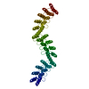

 PDBj
PDBj Assembly
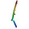


 Mass: 35.453 Da / Num. of mol.: 2 / Source method: obtained synthetically / Formula: Cl
 Mass: 92.094 Da / Num. of mol.: 1 / Source method: obtained synthetically / Formula: C3H8O3
 Mass: 18.015 Da / Num. of mol.: 68 / Source method: isolated from a natural source / Formula: H2O
 Sample preparation
 / Beamline: 23-ID-D / Wavelength: 0.97927 Å
 Processing