 Movie
Movie Controller

[English] 日本語
 Yorodumi
- PDB-2pzt: Crystal structure of Staphylococcal nuclease variant V66Q/P117G/H... -
+ Open data

- Basic information
| Entry | Database: PDB / ID: 2pzt | ||||||
|---|---|---|---|---|---|---|---|
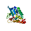
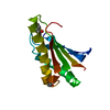
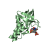
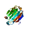
| Title | Crystal structure of Staphylococcal nuclease variant V66Q/P117G/H124L/S128A at 100 K | ||||||
 Components Components | Thermonuclease | ||||||
 Keywords Keywords | HYDROLASE / Staphylococcal nuclease / nuclease / hyperstable variant / internal waters | ||||||
| Function / homology |  Function and homology information Function and homology informationmicrococcal nuclease / 3' overhang single-stranded DNA endonuclease activity / nucleic acid binding / extracellular region / metal ion binding Similarity search - Function | ||||||
| Biological species |   Staphylococcus aureus (bacteria) Staphylococcus aureus (bacteria) | ||||||
| Method |  X-RAY DIFFRACTION / MOLECULAR REPLACEMENT / Resolution: 2.1 Å X-RAY DIFFRACTION / MOLECULAR REPLACEMENT / Resolution: 2.1 Å | ||||||
 Authors Authors | Schlessman, J.L. / Abe, C. / Garcia-Moreno, E.B. | ||||||
 Citation Citation | Journal: Biophys.J. / Year: 2008 Title: Crystallographic study of hydration of an internal cavity in engineered proteins with buried polar or ionizable groups. Authors: Schlessman, J.L. / Abe, C. / Gittis, A. / Karp, D.A. / Dolan, M.A. / Garcia-Moreno, E.B. | ||||||
| History |
|
- Structure visualization
| Structure viewer | Molecule: MolmilJmol/JSmol |
|---|
- Downloads & links
-Download
| PDBx/mmCIF format | 2pzt.cif.gz | 40.2 KB | Display | PDBx/mmCIF format |
|---|---|---|---|---|
| PDB format | pdb2pzt.ent.gz | 27 KB | Display | PDB format |
| PDBx/mmJSON format | 2pzt.json.gz | Tree view | PDBx/mmJSON format | |
| Others |  Other downloads Other downloads |
-Validation report
| Arichive directory | https://data.pdbj.org/pub/pdb/validation_reports/pz/2pztftp://data.pdbj.org/pub/pdb/validation_reports/pz/2pzt | HTTPS FTP |
|---|
-Related structure data
| Related structure data |  2pw5C  2pw7C  2pykC  2pzuC  2pzwC  100kS C: citing same article ( S: Starting model for refinement |
|---|---|
| Similar structure data |


-Links
 PDBj
PDBj- Assembly
| Deposited unit | 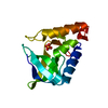
| ||||||||
|---|---|---|---|---|---|---|---|---|---|
| 1 |
| ||||||||
| Unit cell |
|
-Components
| #1: Protein | Mass: 16791.275 Da / Num. of mol.: 1 / Mutation: V66Q, P117G, H124L, S128A Source method: isolated from a genetically manipulated source Source: (gene. exp.) Staphylococcus aureus (bacteria) / Gene: nuc / Plasmid: lambda / Production host: References: UniProt: Q8NXI6, UniProt: A5A523*PLUS, micrococcal nuclease |
|---|---|
| #2: Chemical | ChemComp-PO4 /   Mass: 94.971 Da / Num. of mol.: 1 / Source method: obtained synthetically / Formula: PO4 Mass: 94.971 Da / Num. of mol.: 1 / Source method: obtained synthetically / Formula: PO4 |
| #3: Water | ChemComp-HOH /  Mass: 18.015 Da / Num. of mol.: 69 / Source method: isolated from a natural source / Formula: H2O Mass: 18.015 Da / Num. of mol.: 69 / Source method: isolated from a natural source / Formula: H2O |
-Experimental details
-Experiment
| Experiment | Method: X-RAY DIFFRACTION / Number of used crystals: 1 |
|---|
- Sample preparation
| Crystal | Density Matthews: 2.19 Å3/Da / Density % sol: 43.87 % |
|---|---|
| Crystal grow | Temperature: 277 K / Method: vapor diffusion, hanging drop / pH: 7.8 Details: 37% MPD, 0.025 M potassium phosphate, pH 7.8, VAPOR DIFFUSION, HANGING DROP, temperature 277K |
-Data collection
| Diffraction | Mean temperature: 100 K |
|---|---|
| Diffraction source | Source: SEALED TUBE / Type: OTHER / Wavelength: 1.5418 Å |
| Detector | Type: APEX II CCD / Detector: CCD / Date: Mar 22, 2006 |
| Radiation | Monochromator: GE111 / Protocol: SINGLE WAVELENGTH / Monochromatic (M) / Laue (L): M / Scattering type: x-ray |
| Radiation wavelength | Wavelength: 1.5418 Å / Relative weight: 1 |
| Reflection | Resolution: 2.1→50 Å / Num. all: 8548 / Num. obs: 8548 / % possible obs: 100 % / Observed criterion σ(F): 0 / Observed criterion σ(I): 0 / Redundancy: 12.52 % / Rmerge(I) obs: 0.0236 / Net I/σ(I): 36.73 |
| Reflection shell | Resolution: 2.1→2.2 Å / Redundancy: 8.94 % / Rmerge(I) obs: 0.1334 / Mean I/σ(I) obs: 7.03 / Num. unique all: 1114 / % possible all: 100 |
-Phasing
| Phasing MR | Cor.coef. Fo:Fc: 0.743 / Packing: 0.539
|
|---|
- Processing
| Software |
| |||||||||||||||||||||||||
|---|---|---|---|---|---|---|---|---|---|---|---|---|---|---|---|---|---|---|---|---|---|---|---|---|---|---|
| Refinement | Method to determine structure: MOLECULAR REPLACEMENT Starting model: Staphyloccocal nuclease V66E/P117G/H124L/S128A variant (at 100K), with b-factors set to 20.0 A^2 and residue 66 truncated to Ala, waters removed Resolution: 2.1→50 Å / Isotropic thermal model: isotropic / Cross valid method: THROUGHOUT / σ(F): 0 / Stereochemistry target values: Engh & Huber
| |||||||||||||||||||||||||
| Solvent computation | Bsol: 46.636 Å2 | |||||||||||||||||||||||||
| Displacement parameters | Biso mean: 26.145 Å2
| |||||||||||||||||||||||||
| Refinement step | Cycle: LAST / Resolution: 2.1→50 Å
| |||||||||||||||||||||||||
| Refine LS restraints |
| |||||||||||||||||||||||||
| LS refinement shell | Resolution: 2.1→2.14 Å
| |||||||||||||||||||||||||
| Xplor file |
|
