 Movie
Movie Controller

[English] 日本語
 Yorodumi
- PDB-2pzl: Crystal structure of the Bordetella bronchiseptica enzyme WbmG in... -
+ Open data

- Basic information
| Entry | Database: PDB / ID: 2pzl | ||||||
|---|---|---|---|---|---|---|---|
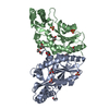
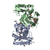
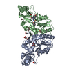
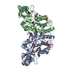
| Title | Crystal structure of the Bordetella bronchiseptica enzyme WbmG in complex with NAD and UDP | ||||||
 Components Components | Putative nucleotide sugar epimerase/ dehydratase | ||||||
 Keywords Keywords | SUGAR BINDING PROTEIN / ROSSMANN FOLD / PROTEIN-NAD COMPLEX / PROTEIN-NUCLEOTIDE COMPLEX | ||||||
| Function / homology |  Function and homology information Function and homology information | ||||||
| Biological species |  Bordetella bronchiseptica (bacteria) Bordetella bronchiseptica (bacteria) | ||||||
| Method |  X-RAY DIFFRACTION / SYNCHROTRON / Single Wavelength / Resolution: 2.39 Å X-RAY DIFFRACTION / SYNCHROTRON / Single Wavelength / Resolution: 2.39 Å | ||||||
 Authors Authors | King, J.D. / Harmer, N.J. / Maskell, D.J. / Blundell, T.L. | ||||||
 Citation Citation | Journal: J.Mol.Biol. / Year: 2007 Title: Predicting protein function from structure--the roles of short-chain dehydrogenase/reductase enzymes in Bordetella O-antigen biosynthesis. Authors: King, J.D. / Harmer, N.J. / Preston, A. / Palmer, C.M. / Rejzek, M. / Field, R.A. / Blundell, T.L. / Maskell, D.J. #1: Journal: Acta Crystallogr.,Sect.F / Year: 2007 Title: Cloning, expression, purification and preliminary crystallographic analysis of the short-chain dehydrogenase enzymes WbmF, WbmG and WbmH from Bordetella bronchiseptica. Authors: Harmer, N.J. / King, J.D. / Palmer, C.M. / Preston, A. / Maskell, D.J. / Blundell, T.L. | ||||||
| History |
|
- Structure visualization
| Structure viewer | Molecule: MolmilJmol/JSmol |
|---|
- Downloads & links
-Download
| PDBx/mmCIF format | 2pzl.cif.gz | 135.2 KB | Display | PDBx/mmCIF format |
|---|---|---|---|---|
| PDB format | pdb2pzl.ent.gz | 104.8 KB | Display | PDB format |
| PDBx/mmJSON format | 2pzl.json.gz | Tree view | PDBx/mmJSON format | |
| Others |  Other downloads Other downloads |
-Validation report
| Arichive directory | https://data.pdbj.org/pub/pdb/validation_reports/pz/2pzlftp://data.pdbj.org/pub/pdb/validation_reports/pz/2pzl | HTTPS FTP |
|---|
-Related structure data
| Related structure data | 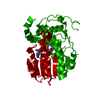 2pzjC 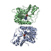 2pzkSC 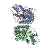 2pzmC 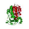 2q1sC 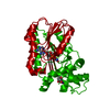 2q1tC 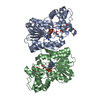 2q1uC 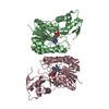 2q1wC S: Starting model for refinement C: citing same article ( |
|---|---|
| Similar structure data |


-Links
 PDBj
PDBj


- Assembly
| Deposited unit | 
| ||||||||||||||||||
|---|---|---|---|---|---|---|---|---|---|---|---|---|---|---|---|---|---|---|---|
| 1 |
| ||||||||||||||||||
| Unit cell |
| ||||||||||||||||||
| Noncrystallographic symmetry (NCS) | NCS domain:
NCS domain segments: Component-ID: 1 / Ens-ID: 1 / Beg auth comp-ID: SER / Beg label comp-ID: SER / End auth comp-ID: SER / End label comp-ID: SER / Refine code: 5 / Auth seq-ID: -1 - 306 / Label seq-ID: 19 - 326
| ||||||||||||||||||
| Details | The biological unit is a dimer, formed from chains A and B. |
-Components
| #1: Protein | Mass: 35287.984 Da / Num. of mol.: 2 Source method: isolated from a genetically manipulated source Source: (gene. exp.) Bordetella bronchiseptica (bacteria) / Gene: BbLPS1.15, wbmG / Plasmid: pET15b / Species (production host): Escherichia coli / Production host: #2: Chemical |   Mass: 663.425 Da / Num. of mol.: 2 / Source method: obtained synthetically / Formula: C21H27N7O14P2 / Comment: NAD*YM Mass: 663.425 Da / Num. of mol.: 2 / Source method: obtained synthetically / Formula: C21H27N7O14P2 / Comment: NAD*YM#3: Chemical |   Type: RNA linking / Mass: 404.161 Da / Num. of mol.: 2 / Source method: obtained synthetically / Formula: C9H14N2O12P2 / Comment: UDP*YM Type: RNA linking / Mass: 404.161 Da / Num. of mol.: 2 / Source method: obtained synthetically / Formula: C9H14N2O12P2 / Comment: UDP*YM#4: Water | ChemComp-HOH / |  Mass: 18.015 Da / Num. of mol.: 154 / Source method: isolated from a natural source / Formula: H2O Mass: 18.015 Da / Num. of mol.: 154 / Source method: isolated from a natural source / Formula: H2O |
|---|
-Experimental details
-Experiment
| Experiment | Method: X-RAY DIFFRACTION / Number of used crystals: 1 |
|---|
- Sample preparation
| Crystal | Density Matthews: 2.51 Å3/Da / Density % sol: 51.01 % |
|---|---|
| Crystal grow | Temperature: 293 K / Method: vapor diffusion, hanging drop / pH: 8.5 Details: 0.1 M Tris, 0.2 M MgCl2, 16-18 % (w/w) PEG 8000. UDP-glucose was added by soaking at 10 mM prior to cryoprotection. pH 8.5, VAPOR DIFFUSION, HANGING DROP, temperature 293K |
-Data collection
| Diffraction | Mean temperature: 100 K |
|---|---|
| Diffraction source | Source: SYNCHROTRON / Site: SRS  / Beamline: PX14.1 / Wavelength: 1.488 Å / Beamline: PX14.1 / Wavelength: 1.488 Å |
| Detector | Type: ADSC QUANTUM 4 / Detector: CCD / Date: Dec 13, 2004 |
| Radiation | Monochromator: Si 111 Channel / Protocol: SINGLE WAVELENGTH / Monochromatic (M) / Laue (L): M / Scattering type: x-ray |
| Radiation wavelength | Wavelength: 1.488 Å / Relative weight: 1 |
| Reflection | Resolution: 2.39→30 Å / Num. all: 27912 / Num. obs: 27549 / % possible obs: 98.7 % / Observed criterion σ(F): 0 / Observed criterion σ(I): 4 / Redundancy: 4.47 % / Rmerge(I) obs: 0.103 / Net I/σ(I): 15 |
| Reflection shell | Resolution: 2.4→2.46 Å / Redundancy: 4.45 % / Rmerge(I) obs: 0.328 / Mean I/σ(I) obs: 3.98 / % possible all: 97.7 |
- Processing
| Software |
| ||||||||||||||||||||||||||||||||||||||||||||||||||||||||||||||||||||||||||||||||||||||||||||||||||||||||||||||||||||||||||||||||||||||||||||||||||||||||||||||||||||||||||
|---|---|---|---|---|---|---|---|---|---|---|---|---|---|---|---|---|---|---|---|---|---|---|---|---|---|---|---|---|---|---|---|---|---|---|---|---|---|---|---|---|---|---|---|---|---|---|---|---|---|---|---|---|---|---|---|---|---|---|---|---|---|---|---|---|---|---|---|---|---|---|---|---|---|---|---|---|---|---|---|---|---|---|---|---|---|---|---|---|---|---|---|---|---|---|---|---|---|---|---|---|---|---|---|---|---|---|---|---|---|---|---|---|---|---|---|---|---|---|---|---|---|---|---|---|---|---|---|---|---|---|---|---|---|---|---|---|---|---|---|---|---|---|---|---|---|---|---|---|---|---|---|---|---|---|---|---|---|---|---|---|---|---|---|---|---|---|---|---|---|---|---|
| Refinement | Method to determine structure: Single Wavelength Starting model: RELATED PDB ENTRY 2PZK WAS SOLVED BY MAD, AND USED AS A STARTING MODEL. Resolution: 2.39→29.97 Å / Cor.coef. Fo:Fc: 0.952 / Cor.coef. Fo:Fc free: 0.918 / SU B: 8.064 / SU ML: 0.189 / Isotropic thermal model: ISOTROPIC / Cross valid method: THROUGHOUT / σ(F): 0 / ESU R: 0.403 / ESU R Free: 0.251 / Stereochemistry target values: MAXIMUM LIKELIHOOD
| ||||||||||||||||||||||||||||||||||||||||||||||||||||||||||||||||||||||||||||||||||||||||||||||||||||||||||||||||||||||||||||||||||||||||||||||||||||||||||||||||||||||||||
| Solvent computation | Ion probe radii: 0.8 Å / Shrinkage radii: 0.8 Å / VDW probe radii: 1.2 Å / Solvent model: MASK | ||||||||||||||||||||||||||||||||||||||||||||||||||||||||||||||||||||||||||||||||||||||||||||||||||||||||||||||||||||||||||||||||||||||||||||||||||||||||||||||||||||||||||
| Displacement parameters | Biso mean: 36.634 Å2
| ||||||||||||||||||||||||||||||||||||||||||||||||||||||||||||||||||||||||||||||||||||||||||||||||||||||||||||||||||||||||||||||||||||||||||||||||||||||||||||||||||||||||||
| Refinement step | Cycle: LAST / Resolution: 2.39→29.97 Å
| ||||||||||||||||||||||||||||||||||||||||||||||||||||||||||||||||||||||||||||||||||||||||||||||||||||||||||||||||||||||||||||||||||||||||||||||||||||||||||||||||||||||||||
| Refine LS restraints |
| ||||||||||||||||||||||||||||||||||||||||||||||||||||||||||||||||||||||||||||||||||||||||||||||||||||||||||||||||||||||||||||||||||||||||||||||||||||||||||||||||||||||||||
| Refine LS restraints NCS | Dom-ID: 1 / Auth asym-ID: A / Ens-ID: 1 / Refine-ID: X-RAY DIFFRACTION
| ||||||||||||||||||||||||||||||||||||||||||||||||||||||||||||||||||||||||||||||||||||||||||||||||||||||||||||||||||||||||||||||||||||||||||||||||||||||||||||||||||||||||||
| LS refinement shell | Resolution: 2.395→2.456 Å / Total num. of bins used: 20
|
