 Movie
Movie Controller

[English] 日本語
 Yorodumi
- PDB-2q1w: Crystal structure of the Bordetella bronchiseptica enzyme WbmH in... -
+ Open data

- Basic information
| Entry | Database: PDB / ID: 2q1w | ||||||
|---|---|---|---|---|---|---|---|
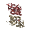
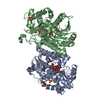
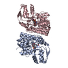
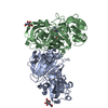
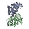
| Title | Crystal structure of the Bordetella bronchiseptica enzyme WbmH in complex with NAD+ | ||||||
 Components Components | Putative nucleotide sugar epimerase/ dehydratase | ||||||
 Keywords Keywords | SUGAR BINDING PROTEIN / ROSSMANN FOLD / PROTEIN-NAD COMPLEX | ||||||
| Function / homology |  Function and homology information Function and homology information | ||||||
| Biological species |  Bordetella bronchiseptica (bacteria) Bordetella bronchiseptica (bacteria) | ||||||
| Method |  X-RAY DIFFRACTION / SYNCHROTRON / MOLECULAR REPLACEMENT / Resolution: 2.19 Å X-RAY DIFFRACTION / SYNCHROTRON / MOLECULAR REPLACEMENT / Resolution: 2.19 Å | ||||||
 Authors Authors | King, J.D. / Harmer, N.J. / Maskell, D.J. / Blundell, T.L. | ||||||
 Citation Citation | Journal: J.Mol.Biol. / Year: 2007 Title: Predicting protein function from structure--the roles of short-chain dehydrogenase/reductase enzymes in Bordetella O-antigen biosynthesis. Authors: King, J.D. / Harmer, N.J. / Preston, A. / Palmer, C.M. / Rejzek, M. / Field, R.A. / Blundell, T.L. / Maskell, D.J. #1: Journal: Acta Crystallogr.,Sect.F / Year: 2007 Title: Cloning, expression, purification and preliminary crystallographic analysis of the short-chain dehydrogenase enzymes WbmF, WbmG and WbmH from Bordetella bronchiseptica. Authors: Harmer, N.J. / King, J.D. / Palmer, C.M. / Preston, A. / Maskell, D.J. / Blundell, T.L. | ||||||
| History |
|
- Structure visualization
| Structure viewer | Molecule: MolmilJmol/JSmol |
|---|
- Downloads & links
-Download
| PDBx/mmCIF format | 2q1w.cif.gz | 192.7 KB | Display | PDBx/mmCIF format |
|---|---|---|---|---|
| PDB format | pdb2q1w.ent.gz | 152 KB | Display | PDB format |
| PDBx/mmJSON format | 2q1w.json.gz | Tree view | PDBx/mmJSON format | |
| Others |  Other downloads Other downloads |
-Validation report
| Arichive directory | https://data.pdbj.org/pub/pdb/validation_reports/q1/2q1wftp://data.pdbj.org/pub/pdb/validation_reports/q1/2q1w | HTTPS FTP |
|---|
-Related structure data
| Related structure data | 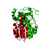 2pzjC 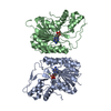 2pzkSC 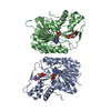 2pzlC 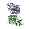 2pzmC 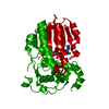 2q1sC 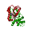 2q1tC 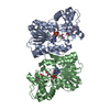 2q1uC S: Starting model for refinement C: citing same article ( |
|---|---|
| Similar structure data |


-Links
 PDBj
PDBj- Assembly
| Deposited unit | 
| ||||||||
|---|---|---|---|---|---|---|---|---|---|
| 1 | 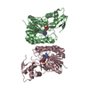
| ||||||||
| 2 | 
| ||||||||
| Unit cell |
| ||||||||
| Details | The biological unit is a dimer. The asymmetric unit contains one complete dimer, formed by chains B and C. A second dimer is formed by chain A, together with a second molecule that is related by rotation around the two-fold axis 0,0,z. |
-Components
| #1: Protein | Mass: 36637.246 Da / Num. of mol.: 3 Source method: isolated from a genetically manipulated source Source: (gene. exp.) Bordetella bronchiseptica (bacteria) / Gene: BbLPS1.14, wbmH / Plasmid: pET15b / Species (production host): Escherichia coli / Production host: #2: Chemical |   Mass: 663.425 Da / Num. of mol.: 3 / Source method: obtained synthetically / Formula: C21H27N7O14P2 / Comment: NAD*YM Mass: 663.425 Da / Num. of mol.: 3 / Source method: obtained synthetically / Formula: C21H27N7O14P2 / Comment: NAD*YM#3: Water | ChemComp-HOH / |  Mass: 18.015 Da / Num. of mol.: 415 / Source method: isolated from a natural source / Formula: H2O Mass: 18.015 Da / Num. of mol.: 415 / Source method: isolated from a natural source / Formula: H2O |
|---|
-Experimental details
-Experiment
| Experiment | Method: X-RAY DIFFRACTION / Number of used crystals: 1 |
|---|
- Sample preparation
| Crystal | Density Matthews: 2.31 Å3/Da / Density % sol: 46.75 % |
|---|---|
| Crystal grow | Temperature: 293 K / Method: vapor diffusion, sitting drop / pH: 7.5 Details: 0.1 M Hepes, 10 % (v/v) isopropanol, 20 % (w/v) PEG 4000, pH 7.5, VAPOR DIFFUSION, SITTING DROP, temperature 293K |
-Data collection
| Diffraction |
| ||||||||||||||||||
|---|---|---|---|---|---|---|---|---|---|---|---|---|---|---|---|---|---|---|---|
| Diffraction source |
| ||||||||||||||||||
| Detector |
| ||||||||||||||||||
| Radiation |
| ||||||||||||||||||
| Radiation wavelength |
| ||||||||||||||||||
| Reflection | Resolution: 2.19→30 Å / Num. all: 53222 / Num. obs: 49975 / % possible obs: 93.9 % / Observed criterion σ(F): 0 / Observed criterion σ(I): 3 / Redundancy: 5.22 % / Rmerge(I) obs: 0.114 / Net I/σ(I): 15.2 | ||||||||||||||||||
| Reflection shell | Resolution: 2.2→2.25 Å / Redundancy: 3.58 % / Rmerge(I) obs: 0.38 / Mean I/σ(I) obs: 3.63 / % possible all: 91.3 |


- Processing
| Software |
| ||||||||||||||||||||||||||||||||||||||||||||||||||||||||||||||||||||||||||||||||||||||||||
|---|---|---|---|---|---|---|---|---|---|---|---|---|---|---|---|---|---|---|---|---|---|---|---|---|---|---|---|---|---|---|---|---|---|---|---|---|---|---|---|---|---|---|---|---|---|---|---|---|---|---|---|---|---|---|---|---|---|---|---|---|---|---|---|---|---|---|---|---|---|---|---|---|---|---|---|---|---|---|---|---|---|---|---|---|---|---|---|---|---|---|---|
| Refinement | Method to determine structure: MOLECULAR REPLACEMENT Starting model: PDB ENTRY 2PZK Resolution: 2.19→30 Å / Cor.coef. Fo:Fc: 0.954 / Cor.coef. Fo:Fc free: 0.936 / SU B: 5.096 / SU ML: 0.132 / Isotropic thermal model: ISOTROPIC / Cross valid method: THROUGHOUT / σ(F): 0 / σ(I): 3.5 / ESU R: 0.257 / ESU R Free: 0.203 / Stereochemistry target values: MAXIMUM LIKELIHOOD
| ||||||||||||||||||||||||||||||||||||||||||||||||||||||||||||||||||||||||||||||||||||||||||
| Solvent computation | Ion probe radii: 0.8 Å / Shrinkage radii: 0.8 Å / VDW probe radii: 1.4 Å / Solvent model: MASK | ||||||||||||||||||||||||||||||||||||||||||||||||||||||||||||||||||||||||||||||||||||||||||
| Displacement parameters | Biso mean: 28.505 Å2
| ||||||||||||||||||||||||||||||||||||||||||||||||||||||||||||||||||||||||||||||||||||||||||
| Refinement step | Cycle: LAST / Resolution: 2.19→30 Å
| ||||||||||||||||||||||||||||||||||||||||||||||||||||||||||||||||||||||||||||||||||||||||||
| Refine LS restraints |
| ||||||||||||||||||||||||||||||||||||||||||||||||||||||||||||||||||||||||||||||||||||||||||
| LS refinement shell | Resolution: 2.185→2.242 Å / Total num. of bins used: 20
|
