 Movie
Movie Controller

[English] 日本語
 Yorodumi
- PDB-2pyf: Crystal Structures of High Affinity Human T-Cell Receptors Bound ... -
+ Open data

- Basic information
| Entry | Database: PDB / ID: 2pyf | ||||||
|---|---|---|---|---|---|---|---|
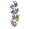
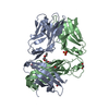
| Title | Crystal Structures of High Affinity Human T-Cell Receptors Bound to pMHC RevealNative Diagonal Binding Geometry Unbound TCR Clone 5-1 | ||||||
 Components Components | (T-Cell Receptor, ...) x 2 | ||||||
 Keywords Keywords | IMMUNE SYSTEM / T-CELL RECEPTOR / CDR3 / PHAGE DISPLAY / MUTANT / HIGH AFFINITY / NY-ESO-1 | ||||||
| Function / homology |  Function and homology information Function and homology informationalpha-beta T cell receptor complex / Translocation of ZAP-70 to Immunological synapse / Phosphorylation of CD3 and TCR zeta chains / alpha-beta T cell activation / Generation of second messenger molecules / Co-inhibition by PD-1 / response to bacterium / Immunoregulatory interactions between a Lymphoid and a non-Lymphoid cell / Downstream TCR signaling / T cell receptor signaling pathway ...alpha-beta T cell receptor complex / Translocation of ZAP-70 to Immunological synapse / Phosphorylation of CD3 and TCR zeta chains / alpha-beta T cell activation / Generation of second messenger molecules / Co-inhibition by PD-1 / response to bacterium / Immunoregulatory interactions between a Lymphoid and a non-Lymphoid cell / Downstream TCR signaling / T cell receptor signaling pathway / adaptive immune response / plasma membrane Similarity search - Function | ||||||
| Biological species |  Homo sapiens (human) Homo sapiens (human) | ||||||
| Method |  X-RAY DIFFRACTION / SYNCHROTRON / MOLECULAR REPLACEMENT / Resolution: 2.2 Å X-RAY DIFFRACTION / SYNCHROTRON / MOLECULAR REPLACEMENT / Resolution: 2.2 Å | ||||||
 Authors Authors | Sami, M. / Rizkallah, P.J. / Dunn, S. / Li, Y. / Moysey, R. / Vuidepot, A. / Baston, E. / Todorov, P. / Molloy, P. / Gao, F. ...Sami, M. / Rizkallah, P.J. / Dunn, S. / Li, Y. / Moysey, R. / Vuidepot, A. / Baston, E. / Todorov, P. / Molloy, P. / Gao, F. / Boulter, J.M. / Jakobsen, B.K. | ||||||
 Citation Citation | Journal: Protein Eng.Des.Sel. / Year: 2007 Title: Crystal structures of high affinity human T-cell receptors bound to peptide major histocompatibility complex reveal native diagonal binding geometry Authors: Sami, M. / Rizkallah, P.J. / Dunn, S. / Molloy, P. / Moysey, R. / Vuidepot, A. / Baston, E. / Todorov, P. / Yi, L. / Gao, F. / Boulter, J.M. / Jakobsen, B.K. | ||||||
| History |
| ||||||
| Remark 999 | Sequence No suitable database reference was found for Chains A and B at time of processing |
- Structure visualization
| Structure viewer | Molecule: MolmilJmol/JSmol |
|---|
- Downloads & links
-Download
| PDBx/mmCIF format | 2pyf.cif.gz | 106.5 KB | Display | PDBx/mmCIF format |
|---|---|---|---|---|
| PDB format | pdb2pyf.ent.gz | 81.4 KB | Display | PDB format |
| PDBx/mmJSON format | 2pyf.json.gz | Tree view | PDBx/mmJSON format | |
| Others |  Other downloads Other downloads |
-Validation report
| Arichive directory | https://data.pdbj.org/pub/pdb/validation_reports/py/2pyfftp://data.pdbj.org/pub/pdb/validation_reports/py/2pyf | HTTPS FTP |
|---|
-Related structure data
| Related structure data |  2p5eC 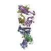 2p5wC 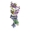 2pyeC 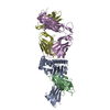 2bnrS C: citing same article ( S: Starting model for refinement |
|---|---|
| Similar structure data |


-Links
 PDBj
PDBj


- Assembly
| Deposited unit | 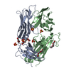
| ||||||||
|---|---|---|---|---|---|---|---|---|---|
| 1 |
| ||||||||
| Unit cell |
|
-Components
-T-Cell Receptor, ... , 2 types, 2 molecules AB
| #1: Protein | Mass: 22437.854 Da / Num. of mol.: 1 Source method: isolated from a genetically manipulated source Source: (gene. exp.) Homo sapiens (human) / Plasmid: PGMT7 / Species (production host): Escherichia coli / Production host:  |
|---|---|
| #2: Protein | Mass: 27020.014 Da / Num. of mol.: 1 Source method: isolated from a genetically manipulated source Source: (gene. exp.) Homo sapiens (human) / Plasmid: PGMT7 / Species (production host): Escherichia coli / Production host: |
-Non-polymers , 4 types, 171 molecules 


| #3: Chemical | ChemComp-SO4 /  Mass: 96.063 Da / Num. of mol.: 6 / Source method: obtained synthetically / Formula: SO4 Mass: 96.063 Da / Num. of mol.: 6 / Source method: obtained synthetically / Formula: SO4#4: Chemical | ChemComp-PG4 / |  Mass: 194.226 Da / Num. of mol.: 1 / Source method: obtained synthetically / Formula: C8H18O5 / Comment: precipitant*YM Mass: 194.226 Da / Num. of mol.: 1 / Source method: obtained synthetically / Formula: C8H18O5 / Comment: precipitant*YM#5: Chemical | ChemComp-PGE /  Mass: 150.173 Da / Num. of mol.: 4 / Source method: obtained synthetically / Formula: C6H14O4 Mass: 150.173 Da / Num. of mol.: 4 / Source method: obtained synthetically / Formula: C6H14O4#6: Water | ChemComp-HOH / | Mass: 18.015 Da / Num. of mol.: 160 / Source method: isolated from a natural source / Formula: H2O |
|---|
-Details
| Has protein modification | Y |
|---|
-Experimental details
-Experiment
| Experiment | Method: X-RAY DIFFRACTION / Number of used crystals: 3 |
|---|
- Sample preparation
| Crystal | Density Matthews: 2.12 Å3/Da / Density % sol: 42.07 % |
|---|---|
| Crystal grow | Temperature: 291 K / Method: vapor diffusion, hanging drop / pH: 7.5 Details: 85 mM Na HEPES buffer pH7.5, 8.5 % iso-propanol, 17% PEG 4000, 15% glycerol, pH 7.50, VAPOR DIFFUSION, HANGING DROP, temperature 291K |
-Data collection
| Diffraction | Mean temperature: 100 K |
|---|---|
| Diffraction source | Source: SYNCHROTRON / Site: SRS  / Beamline: PX14.1 / Wavelength: 1.488 / Beamline: PX14.1 / Wavelength: 1.488 |
| Detector | Type: ADSC QUANTUM 4 / Detector: CCD / Date: Jul 5, 2005 / Details: MIRROR + MONOCHROMATOR |
| Radiation | Monochromator: SI (III) / Protocol: SINGLE WAVELENGTH / Monochromatic (M) / Laue (L): M / Scattering type: x-ray |
| Radiation wavelength | Wavelength: 1.488 Å / Relative weight: 1 |
| Reflection | Resolution: 2.2→30 Å / Num. obs: 20741 / % possible obs: 97 % / Redundancy: 3.2 % / Rmerge(I) obs: 0.139 / Rsym value: 0.139 / Net I/σ(I): 3.9 |
| Reflection shell | Resolution: 2.2→2.32 Å / Redundancy: 3.1 % / Rmerge(I) obs: 0.671 / Mean I/σ(I) obs: 1.3 / Num. measured all: 2196 / Num. unique all: 701 / Rsym value: 0.079 / % possible all: 90.8 |
- Processing
| Software |
| ||||||||||||||||||||||||||||||||||||||||||||||||||||||||||||||||||||||||||||||||||||||||||||||||||||||||||||||||||||||||||||||||||||||||||||||||||||||||||||||||||||||||||
|---|---|---|---|---|---|---|---|---|---|---|---|---|---|---|---|---|---|---|---|---|---|---|---|---|---|---|---|---|---|---|---|---|---|---|---|---|---|---|---|---|---|---|---|---|---|---|---|---|---|---|---|---|---|---|---|---|---|---|---|---|---|---|---|---|---|---|---|---|---|---|---|---|---|---|---|---|---|---|---|---|---|---|---|---|---|---|---|---|---|---|---|---|---|---|---|---|---|---|---|---|---|---|---|---|---|---|---|---|---|---|---|---|---|---|---|---|---|---|---|---|---|---|---|---|---|---|---|---|---|---|---|---|---|---|---|---|---|---|---|---|---|---|---|---|---|---|---|---|---|---|---|---|---|---|---|---|---|---|---|---|---|---|---|---|---|---|---|---|---|---|---|
| Refinement | Method to determine structure: MOLECULAR REPLACEMENT Starting model: 2BNR Resolution: 2.2→30 Å / Cor.coef. Fo:Fc: 0.942 / Cor.coef. Fo:Fc free: 0.879 / SU B: 15.783 / SU ML: 0.221 / TLS residual ADP flag: LIKELY RESIDUAL / Cross valid method: THROUGHOUT / ESU R: 0.426 / ESU R Free: 0.291 / Stereochemistry target values: MAXIMUM LIKELIHOOD / Details: HYDROGENS HAVE BEEN ADDED IN THE RIDING POSITIONS
| ||||||||||||||||||||||||||||||||||||||||||||||||||||||||||||||||||||||||||||||||||||||||||||||||||||||||||||||||||||||||||||||||||||||||||||||||||||||||||||||||||||||||||
| Solvent computation | Ion probe radii: 0.8 Å / Shrinkage radii: 0.8 Å / VDW probe radii: 1.4 Å / Solvent model: MASK | ||||||||||||||||||||||||||||||||||||||||||||||||||||||||||||||||||||||||||||||||||||||||||||||||||||||||||||||||||||||||||||||||||||||||||||||||||||||||||||||||||||||||||
| Displacement parameters | Biso mean: 17.403 Å2
| ||||||||||||||||||||||||||||||||||||||||||||||||||||||||||||||||||||||||||||||||||||||||||||||||||||||||||||||||||||||||||||||||||||||||||||||||||||||||||||||||||||||||||
| Refinement step | Cycle: LAST / Resolution: 2.2→30 Å
| ||||||||||||||||||||||||||||||||||||||||||||||||||||||||||||||||||||||||||||||||||||||||||||||||||||||||||||||||||||||||||||||||||||||||||||||||||||||||||||||||||||||||||
| Refine LS restraints |
| ||||||||||||||||||||||||||||||||||||||||||||||||||||||||||||||||||||||||||||||||||||||||||||||||||||||||||||||||||||||||||||||||||||||||||||||||||||||||||||||||||||||||||
| LS refinement shell | Resolution: 2.2→2.257 Å / Total num. of bins used: 20
| ||||||||||||||||||||||||||||||||||||||||||||||||||||||||||||||||||||||||||||||||||||||||||||||||||||||||||||||||||||||||||||||||||||||||||||||||||||||||||||||||||||||||||
| Refinement TLS params. | Method: refined / Refine-ID: X-RAY DIFFRACTION
| ||||||||||||||||||||||||||||||||||||||||||||||||||||||||||||||||||||||||||||||||||||||||||||||||||||||||||||||||||||||||||||||||||||||||||||||||||||||||||||||||||||||||||
| Refinement TLS group |
|
