 Movie
Movie Controller


+ Open data

- Basic information
| Entry | Database: PDB / ID: 2py0 | ||||||
|---|---|---|---|---|---|---|---|
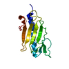
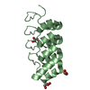
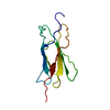
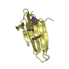
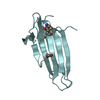
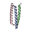
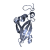
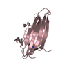
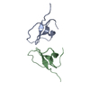
| Title | Crystal structure of Cs1 pilin chimera | ||||||
 Components Components | Fimbrial protein | ||||||
 Keywords Keywords | CELL ADHESION / TYPE IV PILUS / CONSENSUS SEQUENCE / PSEUDOMONAS AERUGINOSA / MONOMERIC PILIN | ||||||
| Function / homology |  Function and homology information Function and homology informationtype IV pilus / type IV pilus-dependent motility / cell adhesion / membrane Similarity search - Function | ||||||
| Biological species |   Pseudomonas aeruginosa (bacteria) Pseudomonas aeruginosa (bacteria) | ||||||
| Method |  X-RAY DIFFRACTION / SYNCHROTRON / MOLECULAR REPLACEMENT / Resolution: 1.35 Å X-RAY DIFFRACTION / SYNCHROTRON / MOLECULAR REPLACEMENT / Resolution: 1.35 Å | ||||||
 Authors Authors | Kao, D.J. / Churchill, M.E. / Hodges, R.S. | ||||||
 Citation Citation | Journal: J.Mol.Biol. / Year: 2007 Title: Animal Protection and Structural Studies of a Consensus Sequence Vaccine Targeting the Receptor Binding Domain of the Type IV Pilus of Pseudomonas aeruginosa. Authors: Kao, D.J. / Churchill, M.E. / Irvin, R.T. / Hodges, R.S. | ||||||
| History |
|
- Structure visualization
| Structure viewer | Molecule: MolmilJmol/JSmol |
|---|
- Downloads & links
-Download
| PDBx/mmCIF format | 2py0.cif.gz | 43.1 KB | Display | PDBx/mmCIF format |
|---|---|---|---|---|
| PDB format | pdb2py0.ent.gz | 28.6 KB | Display | PDB format |
| PDBx/mmJSON format | 2py0.json.gz | Tree view | PDBx/mmJSON format | |
| Others |  Other downloads Other downloads |
-Validation report
| Arichive directory | https://data.pdbj.org/pub/pdb/validation_reports/py/2py0ftp://data.pdbj.org/pub/pdb/validation_reports/py/2py0 | HTTPS FTP |
|---|
-Related structure data
| Related structure data |  1dzoS S: Starting model for refinement |
|---|---|
| Similar structure data |


-Links
 PDBj
PDBj
- Assembly
| Deposited unit | 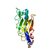
| ||||||||
|---|---|---|---|---|---|---|---|---|---|
| 1 |
| ||||||||
| Unit cell |
|
-Components
| #1: Protein | Mass: 12292.838 Da / Num. of mol.: 1 / Mutation: T136K, E141P Source method: isolated from a genetically manipulated source Source: (gene. exp.) Pseudomonas aeruginosa (bacteria) / Strain: PAK / Gene: pilA, fimA / Plasmid: pRLD / Species (production host): Escherichia coli / Production host: |
|---|---|
| #2: Chemical | ChemComp-EPE /   Mass: 238.305 Da / Num. of mol.: 1 / Source method: obtained synthetically / Formula: C8H18N2O4S / Comment: pH buffer*YM Mass: 238.305 Da / Num. of mol.: 1 / Source method: obtained synthetically / Formula: C8H18N2O4S / Comment: pH buffer*YM |
| #3: Water | ChemComp-HOH /  Mass: 18.015 Da / Num. of mol.: 229 / Source method: isolated from a natural source / Formula: H2O Mass: 18.015 Da / Num. of mol.: 229 / Source method: isolated from a natural source / Formula: H2O |
| Has protein modification | Y |
-Experimental details
-Experiment
| Experiment | Method: X-RAY DIFFRACTION / Number of used crystals: 1 |
|---|
- Sample preparation
| Crystal | Density Matthews: 1.95 Å3/Da / Density % sol: 37.08 % |
|---|---|
| Crystal grow | Temperature: 298 K / Method: vapor diffusion, hanging drop / pH: 8.1 Details: 60% sat. Ammonium sulfate, 0.1 M HEPES., pH 8.1, VAPOR DIFFUSION, HANGING DROP, temperature 298K |
-Data collection
| Diffraction | Mean temperature: 100 K |
|---|---|
| Diffraction source | Source: SYNCHROTRON / Site: APS  / Beamline: 19-ID / Wavelength: 0.97948 Å / Beamline: 19-ID / Wavelength: 0.97948 Å |
| Detector | Type: ADSC QUANTUM 315 / Detector: CCD / Date: Jul 12, 2005 |
| Radiation | Protocol: SINGLE WAVELENGTH / Monochromatic (M) / Laue (L): M / Scattering type: x-ray |
| Radiation wavelength | Wavelength: 0.97948 Å / Relative weight: 1 |
| Reflection | Resolution: 1.35→50 Å / Num. all: 21880 / Num. obs: 21824 / % possible obs: 99.73 % / Redundancy: 7.7 % / Biso Wilson estimate: 14.931 Å2 / Rsym value: 0.051 / Net I/σ(I): 42.8 |
| Reflection shell | Resolution: 1.35→1.385 Å / Redundancy: 7.7 % / Mean I/σ(I) obs: 7 / Num. unique all: 2159 / Rsym value: 0.375 / % possible all: 100 |
-Phasing
| Phasing MR |
|
|---|
- Processing
| Software |
| ||||||||||||||||||||||||||||||||||||||||||||||||||||||||||||||||||||||||||||||||||||||||||
|---|---|---|---|---|---|---|---|---|---|---|---|---|---|---|---|---|---|---|---|---|---|---|---|---|---|---|---|---|---|---|---|---|---|---|---|---|---|---|---|---|---|---|---|---|---|---|---|---|---|---|---|---|---|---|---|---|---|---|---|---|---|---|---|---|---|---|---|---|---|---|---|---|---|---|---|---|---|---|---|---|---|---|---|---|---|---|---|---|---|---|---|
| Refinement | Method to determine structure: MOLECULAR REPLACEMENT Starting model: 1DZO, residues 25-127, backbone atoms only. Resolution: 1.35→25 Å / Cor.coef. Fo:Fc: 0.964 / Cor.coef. Fo:Fc free: 0.957 / SU B: 0.899 / SU ML: 0.038 / Cross valid method: THROUGHOUT / σ(F): 0 / ESU R: 0.064 / ESU R Free: 0.065 / Stereochemistry target values: MAXIMUM LIKELIHOOD / Details: HYDROGENS HAVE BEEN ADDED IN THE RIDING POSITIONS
| ||||||||||||||||||||||||||||||||||||||||||||||||||||||||||||||||||||||||||||||||||||||||||
| Solvent computation | Ion probe radii: 0.8 Å / Shrinkage radii: 0.8 Å / VDW probe radii: 1.2 Å / Solvent model: MASK | ||||||||||||||||||||||||||||||||||||||||||||||||||||||||||||||||||||||||||||||||||||||||||
| Displacement parameters | Biso mean: 14.934 Å2
| ||||||||||||||||||||||||||||||||||||||||||||||||||||||||||||||||||||||||||||||||||||||||||
| Refine analyze | Luzzati coordinate error obs: 0.135 Å | ||||||||||||||||||||||||||||||||||||||||||||||||||||||||||||||||||||||||||||||||||||||||||
| Refinement step | Cycle: LAST / Resolution: 1.35→25 Å
| ||||||||||||||||||||||||||||||||||||||||||||||||||||||||||||||||||||||||||||||||||||||||||
| Refine LS restraints |
| ||||||||||||||||||||||||||||||||||||||||||||||||||||||||||||||||||||||||||||||||||||||||||
| LS refinement shell | Resolution: 1.35→1.385 Å / Total num. of bins used: 20
|
