 Movie
Movie Controller

[English] 日本語
 Yorodumi
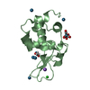
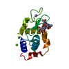
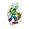
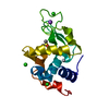
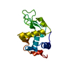
- PDB-2pv1: Crystallographic Structure of SurA first peptidyl-prolyl isomeras... -
+ Open data

- Basic information
| Entry | Database: PDB / ID: 2pv1 | ||||||
|---|---|---|---|---|---|---|---|
| Title | Crystallographic Structure of SurA first peptidyl-prolyl isomerase domain complexed with peptide WEYIPNV | ||||||
 Components Components |
| ||||||
 Keywords Keywords | ISOMERASE / Survival protein A / Peptidyl-prolyl cis-trans isomerase domain / Complex | ||||||
| Function / homology |  Function and homology information Function and homology information: / maintenance of unfolded protein / Gram-negative-bacterium-type cell outer membrane assembly / glycosyltransferase activity / : / peptide binding / peptidylprolyl isomerase / peptidyl-prolyl cis-trans isomerase activity / unfolded protein binding / protein folding ...: / maintenance of unfolded protein / Gram-negative-bacterium-type cell outer membrane assembly / glycosyltransferase activity / : / peptide binding / peptidylprolyl isomerase / peptidyl-prolyl cis-trans isomerase activity / unfolded protein binding / protein folding / outer membrane-bounded periplasmic space / protein stabilization Similarity search - Function | ||||||
| Biological species |  | ||||||
| Method |  X-RAY DIFFRACTION / SYNCHROTRON / MOLECULAR REPLACEMENT / Resolution: 1.3 Å X-RAY DIFFRACTION / SYNCHROTRON / MOLECULAR REPLACEMENT / Resolution: 1.3 Å | ||||||
 Authors Authors | Xu, X. / McKay, D.B. | ||||||
 Citation Citation | Journal: J.Mol.Biol. / Year: 2007 Title: The Periplasmic Bacterial Molecular Chaperone SurA Adapts its Structure to Bind Peptides in Different Conformations to Assert a Sequence Preference for Aromatic Residues. Authors: Xu, X. / Wang, S. / Hu, Y.X. / McKay, D.B. #1: Journal: Structure / Year: 2002Title: Crystallographic structure of SurA, a molecular chaperone that facilitates folding of outer membrane porins Authors: Bitto, E. / McKay, D.B. | ||||||
| History |
|
- Structure visualization
| Structure viewer | Molecule: MolmilJmol/JSmol |
|---|
- Downloads & links
-Download
| PDBx/mmCIF format | 2pv1.cif.gz | 36.6 KB | Display | PDBx/mmCIF format |
|---|---|---|---|---|
| PDB format | pdb2pv1.ent.gz | 23.5 KB | Display | PDB format |
| PDBx/mmJSON format | 2pv1.json.gz | Tree view | PDBx/mmJSON format | |
| Others |  Other downloads Other downloads |
-Validation report
| Summary document | 2pv1_validation.pdf.gz | 422.2 KB | Display | wwPDB validaton report |
|---|---|---|---|---|
| Full document | 2pv1_full_validation.pdf.gz | 424.4 KB | Display | |
| Data in XML | 2pv1_validation.xml.gz | 7.9 KB | Display | |
| Data in CIF | 2pv1_validation.cif.gz | 10.7 KB | Display | |
| Arichive directory | https://data.pdbj.org/pub/pdb/validation_reports/pv/2pv1ftp://data.pdbj.org/pub/pdb/validation_reports/pv/2pv1 | HTTPS FTP |
-Related structure data
| Related structure data |  2pv2C 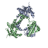 2pv3C  1m5yS S: Starting model for refinement C: citing same article ( |
|---|---|
| Similar structure data |

-Links
 PDBj
PDBj


- Assembly
| Deposited unit | 
| ||||||||
|---|---|---|---|---|---|---|---|---|---|
| 1 |
| ||||||||
| Unit cell |
|
-Components
| #1: Protein | Mass: 10977.320 Da / Num. of mol.: 1 / Fragment: PpiC 1 Source method: isolated from a genetically manipulated source Source: (gene. exp.) |
|---|---|
| #2: Protein/peptide | Mass: 920.019 Da / Num. of mol.: 1 / Source method: obtained synthetically / References: UniProt: Q2RHX9 |
| #3: Water | ChemComp-HOH /  Mass: 18.015 Da / Num. of mol.: 132 / Source method: isolated from a natural source / Formula: H2O Mass: 18.015 Da / Num. of mol.: 132 / Source method: isolated from a natural source / Formula: H2O |
-Experimental details
-Experiment
| Experiment | Method: X-RAY DIFFRACTION / Number of used crystals: 1 |
|---|
- Sample preparation
| Crystal | Density Matthews: 2.21 Å3/Da / Density % sol: 44.36 % |
|---|---|
| Crystal grow | Temperature: 298 K / Method: vapor diffusion, sitting drop / pH: 6.5 Details: 25~28% polyethylene glycol monomethylether 5000, 0.2 M ammonium sulfate, 0.1 M MES buffer, pH 6.5, VAPOR DIFFUSION, SITTING DROP, temperature 298K |
-Data collection
| Diffraction | Mean temperature: 100 K |
|---|---|
| Diffraction source | Source: SYNCHROTRON / Site: SSRL  / Beamline: BL9-2 / Wavelength: 1 Å / Beamline: BL9-2 / Wavelength: 1 Å |
| Detector | Type: MARMOSAIC 325 mm CCD / Detector: CCD / Date: Feb 7, 2007 |
| Radiation | Protocol: SINGLE WAVELENGTH / Monochromatic (M) / Laue (L): M / Scattering type: x-ray |
| Radiation wavelength | Wavelength: 1 Å / Relative weight: 1 |
| Reflection | Resolution: 1.29→50 Å / Num. obs: 26586 / % possible obs: 98.4 % / Redundancy: 4.5 % / Rsym value: 0.034 / Χ2: 2.508 / Net I/σ(I): 39.7 |
| Reflection shell | Resolution: 1.29→1.34 Å / Redundancy: 3.5 % / Num. unique all: 2504 / Rsym value: 0.126 / Χ2: 2.009 / % possible all: 93.8 |
- Processing
| Software |
| ||||||||||||||||||||||||||||
|---|---|---|---|---|---|---|---|---|---|---|---|---|---|---|---|---|---|---|---|---|---|---|---|---|---|---|---|---|---|
| Refinement | Method to determine structure: MOLECULAR REPLACEMENT Starting model: PDB ENTRY 1M5Y Resolution: 1.3→41.05 Å / Cross valid method: THROUGHOUT / σ(F): 0
| ||||||||||||||||||||||||||||
| Solvent computation | Bsol: 44.38 Å2 | ||||||||||||||||||||||||||||
| Displacement parameters | Biso mean: 19.142 Å2
| ||||||||||||||||||||||||||||
| Refine analyze |
| ||||||||||||||||||||||||||||
| Refinement step | Cycle: LAST / Resolution: 1.3→41.05 Å
| ||||||||||||||||||||||||||||
| Refine LS restraints |
| ||||||||||||||||||||||||||||
| LS refinement shell | Resolution: 1.3→1.38 Å / Rfactor Rfree error: 0.014
| ||||||||||||||||||||||||||||
| Xplor file |
|
