 Movie
Movie Controller

[English] 日本語
 Yorodumi
- PDB-2pua: CRYSTAL STRUCTURE OF THE LACI FAMILY MEMBER, PURR, BOUND TO DNA: ... -
+ Open data

- Basic information
| Entry | Database: PDB / ID: 2pua | ||||||
|---|---|---|---|---|---|---|---|
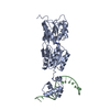
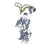
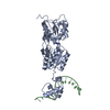
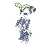
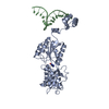
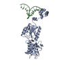
| Title | CRYSTAL STRUCTURE OF THE LACI FAMILY MEMBER, PURR, BOUND TO DNA: MINOR GROOVE BINDING BY ALPHA HELICES | ||||||
 Components Components |
| ||||||
 Keywords Keywords | TRANSCRIPTION/DNA / COMPLEX (DNA-BINDING PROTEIN-DNA) / DNA-BINDING REGULATORY PROTEIN / TRANSCRIPTION-DNA COMPLEX | ||||||
| Function / homology |  Function and homology information Function and homology informationguanine binding / negative regulation of purine nucleotide biosynthetic process / purine nucleotide biosynthetic process / DNA-binding transcription repressor activity / transcription cis-regulatory region binding / DNA-binding transcription factor activity / negative regulation of DNA-templated transcription / regulation of DNA-templated transcription / protein homodimerization activity / cytosol Similarity search - Function | ||||||
| Biological species |  synthetic construct (others) | ||||||
| Method |  X-RAY DIFFRACTION / Resolution: 2.9 Å X-RAY DIFFRACTION / Resolution: 2.9 Å | ||||||
 Authors Authors | Schumacher, R.G. / Choi, K.Y. / Zalkin, H. / Brennan, M.A. | ||||||
 Citation Citation | Journal: Science / Year: 1994 Title: Crystal structure of LacI member, PurR, bound to DNA: minor groove binding by alpha helices. Authors: Schumacher, M.A. / Choi, K.Y. / Zalkin, H. / Brennan, R.G. #1: Journal: Biochemistry / Year: 1998Title: Structure-Based Redesign of Corepressor Specificity of the Escherichia Coli Purine Repressor by Substitution of Residue 190 Authors: Lu, F. / Schumacher, M.A. / Arvidson, D.N. / Haldimann, A. / Wanner, B.L. / Zalkin, H. / Brennan, R.G. | ||||||
| History |
|
- Structure visualization
| Structure viewer | Molecule: MolmilJmol/JSmol |
|---|
- Downloads & links
-Download
| PDBx/mmCIF format | 2pua.cif.gz | 92.7 KB | Display | PDBx/mmCIF format |
|---|---|---|---|---|
| PDB format | pdb2pua.ent.gz | 66.5 KB | Display | PDB format |
| PDBx/mmJSON format | 2pua.json.gz | Tree view | PDBx/mmJSON format | |
| Others |  Other downloads Other downloads |
-Validation report
| Arichive directory | https://data.pdbj.org/pub/pdb/validation_reports/pu/2puaftp://data.pdbj.org/pub/pdb/validation_reports/pu/2pua | HTTPS FTP |
|---|
-Related structure data


-Links
 PDBj
PDBj


- Assembly
| Deposited unit | 
| ||||||||
|---|---|---|---|---|---|---|---|---|---|
| 1 | 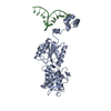
| ||||||||
| Unit cell |
| ||||||||
| Details | THE COMPLEX LIES ON A CRYSTALLOGRAPHIC TWO-FOLD AXIS, AND ONLY ONE MONOMER-DNA HALF-SITE CONSTITUTES THE ASYMMETRIC UNIT. THE COORDINATES COMPRISE ONE REPRESSOR MONOMER AND ONE DNA STRAND FOR THE ENTIRE SITE. THE FULL COMPLEX CAN BE CONSTRUCTED BY GENERATING THE SECOND HALF USING THE CRYSTALLOGRAPHIC SYMMETRY OPERATION (X, -Y, -Z). |
-Components
| #1: DNA chain | Mass: 5202.384 Da / Num. of mol.: 1 Source method: isolated from a genetically manipulated source Source: (synth.) synthetic construct (others) |
|---|---|
| #2: Protein | Mass: 38005.480 Da / Num. of mol.: 1 / Mutation: R190A Source method: isolated from a genetically manipulated source Source: (gene. exp.) |
| #3: Chemical | ChemComp-6MP /   Mass: 134.139 Da / Num. of mol.: 1 / Source method: obtained synthetically / Formula: C6H6N4 Mass: 134.139 Da / Num. of mol.: 1 / Source method: obtained synthetically / Formula: C6H6N4 |
| #4: Water | ChemComp-HOH /  Mass: 18.015 Da / Num. of mol.: 59 / Source method: isolated from a natural source / Formula: H2O Mass: 18.015 Da / Num. of mol.: 59 / Source method: isolated from a natural source / Formula: H2O |
| Compound details | THE HYDROGEN BONDS FROM ARG 190 TO THE O6 EXOCYCLIC ATOM IS WHAT DETERMINES PURR'S SPECIFICITY FOR ...THE HYDROGEN BONDS FROM ARG 190 TO THE O6 EXOCYCLIC ATOM IS WHAT DETERMINES |
-Experimental details
-Experiment
| Experiment | Method: X-RAY DIFFRACTION / Number of used crystals: 1 |
|---|
- Sample preparation
| Crystal | Density Matthews: 3.94 Å3/Da / Density % sol: 68.76 % | ||||||||||||||||||||||||||||||||||||||||||||||||||||||||||||||||||||||||||||||||||||
|---|---|---|---|---|---|---|---|---|---|---|---|---|---|---|---|---|---|---|---|---|---|---|---|---|---|---|---|---|---|---|---|---|---|---|---|---|---|---|---|---|---|---|---|---|---|---|---|---|---|---|---|---|---|---|---|---|---|---|---|---|---|---|---|---|---|---|---|---|---|---|---|---|---|---|---|---|---|---|---|---|---|---|---|---|---|
| Crystal grow | Temperature: 293 K / Method: vapor diffusion, hanging drop / pH: 7.4 Details: pH 7.40, VAPOR DIFFUSION, HANGING DROP, temperature 293.00K | ||||||||||||||||||||||||||||||||||||||||||||||||||||||||||||||||||||||||||||||||||||
| Components of the solutions |
| ||||||||||||||||||||||||||||||||||||||||||||||||||||||||||||||||||||||||||||||||||||
| Crystal grow | *PLUS Details: Schumacher, M.A., (1994) J.Mol.Biol, 242, 302. / pH: 7.4 | ||||||||||||||||||||||||||||||||||||||||||||||||||||||||||||||||||||||||||||||||||||
| Components of the solutions | *PLUS
|
-Data collection
| Diffraction | Mean temperature: 293 K |
|---|---|
| Diffraction source | Source: ROTATING ANODE / Type: RIGAKU RU200 / Wavelength: 1.5418 |
| Detector | Type: SDMS / Detector: AREA DETECTOR |
| Radiation | Scattering type: x-ray |
| Radiation wavelength | Wavelength: 1.5418 Å / Relative weight: 1 |
| Reflection | Highest resolution: 2.9 Å / Num. obs: 14911 / % possible obs: 99 % / Redundancy: 3.43 % / Rmerge(I) obs: 0.09 / Net I/σ(I): 5.7 |
- Processing
| Software |
| ||||||||||||||||||||||||||||||||||||||||||||||||||
|---|---|---|---|---|---|---|---|---|---|---|---|---|---|---|---|---|---|---|---|---|---|---|---|---|---|---|---|---|---|---|---|---|---|---|---|---|---|---|---|---|---|---|---|---|---|---|---|---|---|---|---|
| Refinement | Resolution: 2.9→10 Å / Isotropic thermal model: ISOTROPIC / σ(F): 1
| ||||||||||||||||||||||||||||||||||||||||||||||||||
| Refinement step | Cycle: LAST / Resolution: 2.9→10 Å
| ||||||||||||||||||||||||||||||||||||||||||||||||||
| Refine LS restraints |
| ||||||||||||||||||||||||||||||||||||||||||||||||||
| Software | *PLUS Name: TNT / Classification: refinement | ||||||||||||||||||||||||||||||||||||||||||||||||||
| Refinement | *PLUS Highest resolution: 2.9 Å / Lowest resolution: 10 Å / Num. reflection all: 18818 / σ(F): 1 | ||||||||||||||||||||||||||||||||||||||||||||||||||
| Solvent computation | *PLUS | ||||||||||||||||||||||||||||||||||||||||||||||||||
| Displacement parameters | *PLUS | ||||||||||||||||||||||||||||||||||||||||||||||||||
| Refine LS restraints | *PLUS
|
