 Movie
Movie Controller


+ Open data

- Basic information
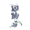
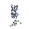
| Entry | Database: PDB / ID: 1qp4 | ||||||
|---|---|---|---|---|---|---|---|
| Title | PURINE REPRESSOR-HYPOXANTHINE-PALINDROMIC OPERATOR COMPLEX | ||||||
 Components Components |
| ||||||
 Keywords Keywords | TRANSCRIPTION/DNA / TRANSCRIPTION REGULATION / DNA-BINDING / REPRESSOR / PURINE BIOSYNTHESIS / COMPLEX (DNA-BINDING PROTEIN-DNA) / TRANSCRIPTION-DNA COMPLEX | ||||||
| Function / homology |  Function and homology information Function and homology informationguanine binding / negative regulation of purine nucleotide biosynthetic process / purine nucleotide biosynthetic process / DNA-binding transcription repressor activity / transcription cis-regulatory region binding / DNA-binding transcription factor activity / negative regulation of DNA-templated transcription / regulation of DNA-templated transcription / DNA-templated transcription / protein homodimerization activity / cytosol Similarity search - Function | ||||||
| Biological species |  | ||||||
| Method |  X-RAY DIFFRACTION / Resolution: 3 Å X-RAY DIFFRACTION / Resolution: 3 Å | ||||||
 Authors Authors | Glasfeld, A. / Koehler, A.N. / Schumacher, M.A. / Brennan, R.G. | ||||||
 Citation Citation | Journal: J.Mol.Biol. / Year: 1999 Title: The role of lysine 55 in determining the specificity of the purine repressor for its operators through minor groove interactions. Authors: Glasfeld, A. / Koehler, A.N. / Schumacher, M.A. / Brennan, R.G. #1: Journal: Science / Year: 1994Title: Crystal Structure of LacI Member, PurR, Bound to DNA: Minor Groove Binding by Alpha Helices Authors: Schumacher, M.A. / Choi, K.Y. / Zalkin, H. / Brennan, R.G. | ||||||
| History |
|
- Structure visualization
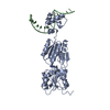
| Structure viewer | Molecule: MolmilJmol/JSmol |
|---|
- Downloads & links
-Download
| PDBx/mmCIF format | 1qp4.cif.gz | 90.3 KB | Display | PDBx/mmCIF format |
|---|---|---|---|---|
| PDB format | pdb1qp4.ent.gz | 65.2 KB | Display | PDB format |
| PDBx/mmJSON format | 1qp4.json.gz | Tree view | PDBx/mmJSON format | |
| Others |  Other downloads Other downloads |
-Validation report
| Arichive directory | https://data.pdbj.org/pub/pdb/validation_reports/qp/1qp4ftp://data.pdbj.org/pub/pdb/validation_reports/qp/1qp4 | HTTPS FTP |
|---|
-Related structure data


-Links
 PDBj
PDBj


- Assembly
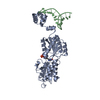
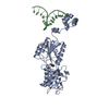
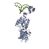
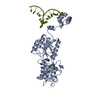
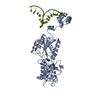
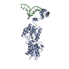
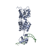
| Deposited unit | 
| ||||||||
|---|---|---|---|---|---|---|---|---|---|
| 1 | 
| ||||||||
| Unit cell |
|
-Components
| #1: DNA chain | Mass: 5202.384 Da / Num. of mol.: 1 / Source method: obtained synthetically |
|---|---|
| #2: Protein | Mass: 38091.594 Da / Num. of mol.: 1 Source method: isolated from a genetically manipulated source Source: (gene. exp.) |
| #3: Chemical | ChemComp-HPA /   Mass: 136.111 Da / Num. of mol.: 1 / Source method: obtained synthetically / Formula: C5H4N4O Mass: 136.111 Da / Num. of mol.: 1 / Source method: obtained synthetically / Formula: C5H4N4O |
| #4: Water | ChemComp-HOH /  Mass: 18.015 Da / Num. of mol.: 2 / Source method: isolated from a natural source / Formula: H2O Mass: 18.015 Da / Num. of mol.: 2 / Source method: isolated from a natural source / Formula: H2O |
-Experimental details
-Experiment
| Experiment | Method: X-RAY DIFFRACTION / Number of used crystals: 1 |
|---|
- Sample preparation
| Crystal | Density Matthews: 3.92 Å3/Da / Density % sol: 68.63 % | ||||||||||||||||||||||||||||||||||||||||||||||||||||||||||||||||||||||
|---|---|---|---|---|---|---|---|---|---|---|---|---|---|---|---|---|---|---|---|---|---|---|---|---|---|---|---|---|---|---|---|---|---|---|---|---|---|---|---|---|---|---|---|---|---|---|---|---|---|---|---|---|---|---|---|---|---|---|---|---|---|---|---|---|---|---|---|---|---|---|---|
| Crystal grow | Temperature: 298 K / Method: vapor diffusion, hanging drop / pH: 7.5 Details: PEG 4000, (NH4)2SO4, [CO(NH3)6]CL3, NA2SO4, pH 7.5, VAPOR DIFFUSION, HANGING DROP, temperature 298K | ||||||||||||||||||||||||||||||||||||||||||||||||||||||||||||||||||||||
| Components of the solutions |
| ||||||||||||||||||||||||||||||||||||||||||||||||||||||||||||||||||||||
| Crystal grow | *PLUS | ||||||||||||||||||||||||||||||||||||||||||||||||||||||||||||||||||||||
| Components of the solutions | *PLUS
|
-Data collection
| Diffraction | Mean temperature: 298 K |
|---|---|
| Diffraction source | Source: ROTATING ANODE / Type: RIGAKU / Wavelength: 1.5418 |
| Detector | Type: UCSD MARK III / Detector: AREA DETECTOR / Date: Feb 14, 1996 |
| Radiation | Protocol: SINGLE WAVELENGTH / Monochromatic (M) / Laue (L): M / Scattering type: x-ray |
| Radiation wavelength | Wavelength: 1.5418 Å / Relative weight: 1 |
| Reflection | Resolution: 3→10 Å / Num. obs: 13547 / % possible obs: 99 % / Observed criterion σ(I): 2 / Redundancy: 4.3 % / Biso Wilson estimate: 41 Å2 / Rsym value: 7.97 / Net I/σ(I): 9.32 |
| Reflection shell | Resolution: 3→3.23 Å / Redundancy: 1.8 % / % possible all: 89.6 |
| Reflection | *PLUS Num. measured all: 58105 / Rmerge(I) obs: 0.0797 |
| Reflection shell | *PLUS % possible obs: 89.6 % |
- Processing
| Software |
| ||||||||||||||||||||||||||||||||||||||||
|---|---|---|---|---|---|---|---|---|---|---|---|---|---|---|---|---|---|---|---|---|---|---|---|---|---|---|---|---|---|---|---|---|---|---|---|---|---|---|---|---|---|
| Refinement | Resolution: 3→10 Å / σ(I): 2 Stereochemistry target values: DNA BASE GEOMETRIES TAKEN FROM CLOWNEY ET AL. (1996) J. AM. CHEM. SOC., VOL. 118, PP. 509-518. SUGAR PHOSPHATE GEOMETRIES TAKEN FROM GELBIN ET AL. (1996) J. AM. CHEM. ...Stereochemistry target values: DNA BASE GEOMETRIES TAKEN FROM CLOWNEY ET AL. (1996) J. AM. CHEM. SOC., VOL. 118, PP. 509-518. SUGAR PHOSPHATE GEOMETRIES TAKEN FROM GELBIN ET AL. (1996) J. AM. CHEM. SOC., VOL. 118, PP. 519-529.
| ||||||||||||||||||||||||||||||||||||||||
| Refinement step | Cycle: LAST / Resolution: 3→10 Å
| ||||||||||||||||||||||||||||||||||||||||
| Refine LS restraints |
|
