 Movie
Movie Controller

[English] 日本語
 Yorodumi
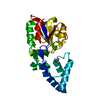
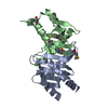
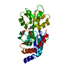
- PDB-2ptt: Structure of NK cell receptor 2B4 (CD244) bound to its ligand CD48 -
+ Open data

- Basic information
| Entry | Database: PDB / ID: 2ptt | ||||||
|---|---|---|---|---|---|---|---|
| Title | Structure of NK cell receptor 2B4 (CD244) bound to its ligand CD48 | ||||||
 Components Components |
| ||||||
 Keywords Keywords | IMMUNE SYSTEM / 2B4 / CD244 / CD48 / NK cell receptor | ||||||
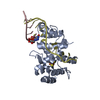
| Function / homology |  Function and homology information Function and homology informationnatural killer cell activation involved in immune response / myeloid dendritic cell activation / mast cell activation / Cell surface interactions at the vascular wall / natural killer cell activation / positive regulation of CD8-positive, alpha-beta T cell proliferation / positive regulation of natural killer cell proliferation / MHC class I protein binding / T cell activation / adaptive immune response ...natural killer cell activation involved in immune response / myeloid dendritic cell activation / mast cell activation / Cell surface interactions at the vascular wall / natural killer cell activation / positive regulation of CD8-positive, alpha-beta T cell proliferation / positive regulation of natural killer cell proliferation / MHC class I protein binding / T cell activation / adaptive immune response / immune response / membrane raft / receptor ligand activity / external side of plasma membrane / signal transduction / protein-containing complex / extracellular region Similarity search - Function | ||||||
| Biological species |  | ||||||
| Method |  X-RAY DIFFRACTION / SYNCHROTRON / MOLECULAR REPLACEMENT / Resolution: 1.63 Å X-RAY DIFFRACTION / SYNCHROTRON / MOLECULAR REPLACEMENT / Resolution: 1.63 Å | ||||||
 Authors Authors | Deng, L. / Velikovsky, C.A. / Mariuzza, R.A. | ||||||
 Citation Citation | Journal: Immunity / Year: 2007 Title: Structure of natural killer receptor 2B4 bound to CD48 reveals basis for heterophilic recognition in signaling lymphocyte activation molecule family. Authors: Velikovsky, C.A. / Deng, L. / Chlewicki, L.K. / Fernandez, M.M. / Kumar, V. / Mariuzza, R.A. | ||||||
| History |
|
- Structure visualization
| Structure viewer | Molecule: MolmilJmol/JSmol |
|---|
- Downloads & links
-Download
| PDBx/mmCIF format | 2ptt.cif.gz | 61.7 KB | Display | PDBx/mmCIF format |
|---|---|---|---|---|
| PDB format | pdb2ptt.ent.gz | 44.2 KB | Display | PDB format |
| PDBx/mmJSON format | 2ptt.json.gz | Tree view | PDBx/mmJSON format | |
| Others |  Other downloads Other downloads |
-Validation report
| Arichive directory | https://data.pdbj.org/pub/pdb/validation_reports/pt/2pttftp://data.pdbj.org/pub/pdb/validation_reports/pt/2ptt | HTTPS FTP |
|---|
-Related structure data
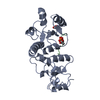
| Related structure data | 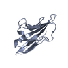 2ptuC  2ptvC  1hngS C: citing same article ( S: Starting model for refinement |
|---|---|
| Similar structure data |


-Links
 PDBj
PDBj


- Assembly
| Deposited unit | 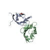
| |||||||||
|---|---|---|---|---|---|---|---|---|---|---|
| 1 |
| |||||||||
| Unit cell |
| |||||||||
| Components on special symmetry positions |
|
-Components
| #1: Protein | Mass: 12940.734 Da / Num. of mol.: 1 / Fragment: Ig-like C2-type1, D1, 2B4-binding domain / Mutation: T34Y, K52R Source method: isolated from a genetically manipulated source Source: (gene. exp.)  |
|---|---|
| #2: Protein | Mass: 12526.927 Da / Num. of mol.: 1 / Fragment: IG-LIKE 1, D1, CD48-binding domain Source method: isolated from a genetically manipulated source Source: (gene. exp.) |
| #3: Chemical | ChemComp-SO4 /   Mass: 96.063 Da / Num. of mol.: 1 / Source method: obtained synthetically / Formula: SO4 Mass: 96.063 Da / Num. of mol.: 1 / Source method: obtained synthetically / Formula: SO4 |
| #4: Water | ChemComp-HOH /  Mass: 18.015 Da / Num. of mol.: 213 / Source method: isolated from a natural source / Formula: H2O Mass: 18.015 Da / Num. of mol.: 213 / Source method: isolated from a natural source / Formula: H2O |
| Has protein modification | Y |
-Experimental details
-Experiment
| Experiment | Method: X-RAY DIFFRACTION / Number of used crystals: 1 |
|---|
- Sample preparation
| Crystal | Density Matthews: 2.25 Å3/Da / Density % sol: 45.25 % |
|---|---|
| Crystal grow | Temperature: 277 K / Method: vapor diffusion, hanging drop / pH: 6.5 Details: 1.6 M ammonium sulfate, 0.1 mM MES, 10% dioxane, pH 6.5, VAPOR DIFFUSION, HANGING DROP, temperature 277K |
-Data collection
| Diffraction | Mean temperature: 100 K |
|---|---|
| Diffraction source | Source: SYNCHROTRON / Site: NSLS  / Beamline: X29A / Wavelength: 1 Å / Beamline: X29A / Wavelength: 1 Å |
| Detector | Type: ADSC QUANTUM 315 / Detector: CCD / Date: Jun 1, 2006 |
| Radiation | Protocol: SINGLE WAVELENGTH / Monochromatic (M) / Laue (L): M / Scattering type: x-ray |
| Radiation wavelength | Wavelength: 1 Å / Relative weight: 1 |
| Reflection | Resolution: 1.63→50 Å / Num. obs: 28061 / % possible obs: 99.1 % / Redundancy: 6.8 % / Biso Wilson estimate: 19.9 Å2 / Rmerge(I) obs: 0.055 / Net I/σ(I): 53.6 |
| Reflection shell | Resolution: 1.63→1.69 Å / Redundancy: 5.9 % / Rmerge(I) obs: 0.18 / Mean I/σ(I) obs: 8 / Num. unique all: 2733 / % possible all: 97.4 |
- Processing
| Software |
| ||||||||||||||||||||||||||||||||||||||||||||||||||||||||||||||||||||||||||||||||||||||||||||||||||||||||||||||||||||||||||||||||||||||||||||||||||||||||||||||||||||||||||
|---|---|---|---|---|---|---|---|---|---|---|---|---|---|---|---|---|---|---|---|---|---|---|---|---|---|---|---|---|---|---|---|---|---|---|---|---|---|---|---|---|---|---|---|---|---|---|---|---|---|---|---|---|---|---|---|---|---|---|---|---|---|---|---|---|---|---|---|---|---|---|---|---|---|---|---|---|---|---|---|---|---|---|---|---|---|---|---|---|---|---|---|---|---|---|---|---|---|---|---|---|---|---|---|---|---|---|---|---|---|---|---|---|---|---|---|---|---|---|---|---|---|---|---|---|---|---|---|---|---|---|---|---|---|---|---|---|---|---|---|---|---|---|---|---|---|---|---|---|---|---|---|---|---|---|---|---|---|---|---|---|---|---|---|---|---|---|---|---|---|---|---|
| Refinement | Method to determine structure: MOLECULAR REPLACEMENT Starting model: PDB ENTRY 1HNG Resolution: 1.63→50 Å / Cor.coef. Fo:Fc: 0.954 / Cor.coef. Fo:Fc free: 0.93 / SU B: 1.712 / SU ML: 0.062 / Cross valid method: THROUGHOUT / ESU R: 0.101 / ESU R Free: 0.109 / Stereochemistry target values: MAXIMUM LIKELIHOOD
| ||||||||||||||||||||||||||||||||||||||||||||||||||||||||||||||||||||||||||||||||||||||||||||||||||||||||||||||||||||||||||||||||||||||||||||||||||||||||||||||||||||||||||
| Solvent computation | Ion probe radii: 0.8 Å / Shrinkage radii: 0.8 Å / VDW probe radii: 1.2 Å / Solvent model: MASK | ||||||||||||||||||||||||||||||||||||||||||||||||||||||||||||||||||||||||||||||||||||||||||||||||||||||||||||||||||||||||||||||||||||||||||||||||||||||||||||||||||||||||||
| Displacement parameters | Biso mean: 21.428 Å2
| ||||||||||||||||||||||||||||||||||||||||||||||||||||||||||||||||||||||||||||||||||||||||||||||||||||||||||||||||||||||||||||||||||||||||||||||||||||||||||||||||||||||||||
| Refinement step | Cycle: LAST / Resolution: 1.63→50 Å
| ||||||||||||||||||||||||||||||||||||||||||||||||||||||||||||||||||||||||||||||||||||||||||||||||||||||||||||||||||||||||||||||||||||||||||||||||||||||||||||||||||||||||||
| Refine LS restraints |
| ||||||||||||||||||||||||||||||||||||||||||||||||||||||||||||||||||||||||||||||||||||||||||||||||||||||||||||||||||||||||||||||||||||||||||||||||||||||||||||||||||||||||||
| LS refinement shell | Resolution: 1.631→1.674 Å / Total num. of bins used: 20
|
