 Movie
Movie Controller


+ Open data

- Basic information
| Entry | Database: PDB / ID: 2pth | ||||||
|---|---|---|---|---|---|---|---|
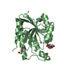
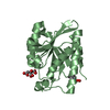
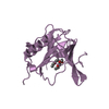
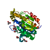
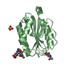
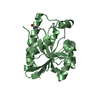
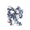
| Title | PEPTIDYL-TRNA HYDROLASE FROM ESCHERICHIA COLI | ||||||
 Components Components | PEPTIDYL-TRNA HYDROLASE | ||||||
 Keywords Keywords | HYDROLASE / PEPTIDYL-TRNA | ||||||
| Function / homology |  Function and homology information Function and homology informationpeptidyl-tRNA hydrolase / peptidyl-tRNA hydrolase activity / protein quality control for misfolded or incompletely synthesized proteins / rescue of stalled ribosome / tRNA binding / cytoplasm Similarity search - Function | ||||||
| Biological species |  | ||||||
| Method |  X-RAY DIFFRACTION / SYNCHROTRON / MIRS / Resolution: 1.2 Å X-RAY DIFFRACTION / SYNCHROTRON / MIRS / Resolution: 1.2 Å | ||||||
 Authors Authors | Schmitt, E. / Mechulam, Y. / Fromant, M. / Plateau, P. / Blanquet, S. | ||||||
 Citation Citation | Journal: EMBO J. / Year: 1997 Title: Crystal structure at 1.2 A resolution and active site mapping of Escherichia coli peptidyl-tRNA hydrolase. Authors: Schmitt, E. / Mechulam, Y. / Fromant, M. / Plateau, P. / Blanquet, S. | ||||||
| History |
|
- Structure visualization
| Structure viewer | Molecule: MolmilJmol/JSmol |
|---|
- Downloads & links
-Download
| PDBx/mmCIF format | 2pth.cif.gz | 52.5 KB | Display | PDBx/mmCIF format |
|---|---|---|---|---|
| PDB format | pdb2pth.ent.gz | 38.4 KB | Display | PDB format |
| PDBx/mmJSON format | 2pth.json.gz | Tree view | PDBx/mmJSON format | |
| Others |  Other downloads Other downloads |
-Validation report
| Summary document | 2pth_validation.pdf.gz | 364.8 KB | Display | wwPDB validaton report |
|---|---|---|---|---|
| Full document | 2pth_full_validation.pdf.gz | 366.5 KB | Display | |
| Data in XML | 2pth_validation.xml.gz | 5.3 KB | Display | |
| Data in CIF | 2pth_validation.cif.gz | 8.5 KB | Display | |
| Arichive directory | https://data.pdbj.org/pub/pdb/validation_reports/pt/2pthftp://data.pdbj.org/pub/pdb/validation_reports/pt/2pth | HTTPS FTP |
-Related structure data
| Similar structure data |
|---|


-Links
 PDBj
PDBj- Assembly
| Deposited unit | 
| ||||||||
|---|---|---|---|---|---|---|---|---|---|
| 1 |
| ||||||||
| Unit cell |
|
-Components
| #1: Protein | Mass: 20982.115 Da / Num. of mol.: 1 Source method: isolated from a genetically manipulated source Source: (gene. exp.) |
|---|---|
| #2: Water | ChemComp-HOH /  Mass: 18.015 Da / Num. of mol.: 181 / Source method: isolated from a natural source / Formula: H2O Mass: 18.015 Da / Num. of mol.: 181 / Source method: isolated from a natural source / Formula: H2O |
-Experimental details
-Experiment
| Experiment | Method: X-RAY DIFFRACTION / Number of used crystals: 1 |
|---|
- Sample preparation
| Crystal | Density Matthews: 2.24 Å3/Da / Density % sol: 45.05 % | |||||||||||||||||||||||||
|---|---|---|---|---|---|---|---|---|---|---|---|---|---|---|---|---|---|---|---|---|---|---|---|---|---|---|
| Crystal grow | pH: 7.5 / Details: 10% PEG6000,12% ISOPROPANOL, 0,1 M TRIS-CL PH7.5 | |||||||||||||||||||||||||
| Crystal grow | *PLUS Temperature: 6 ℃ / Method: vapor diffusion, hanging dropDetails: Schmitt, E., (1997) Proteins.Struct.Funct.Genet., 28, 135. | |||||||||||||||||||||||||
| Components of the solutions | *PLUS
|
-Data collection
| Diffraction | Mean temperature: 273 K |
|---|---|
| Diffraction source | Source: SYNCHROTRON / Site: LURE  / Beamline: DW32 / Wavelength: 0.994 / Beamline: DW32 / Wavelength: 0.994 |
| Detector | Type: MARRESEARCH / Detector: IMAGE PLATE / Date: Feb 1, 1996 |
| Radiation | Monochromatic (M) / Laue (L): M / Scattering type: x-ray |
| Radiation wavelength | Wavelength: 0.994 Å / Relative weight: 1 |
| Reflection | Resolution: 1.2→28 Å / Num. obs: 55575 / % possible obs: 93.9 % / Observed criterion σ(I): 0 / Redundancy: 4.2 % / Biso Wilson estimate: 12.7 Å2 / Rsym value: 0.046 / Net I/σ(I): 8.3 |
| Reflection shell | Resolution: 1.2→1.23 Å / Redundancy: 3.7 % / Mean I/σ(I) obs: 4.6 / Rsym value: 0.15 / % possible all: 85.3 |
| Reflection | *PLUS Rmerge(I) obs: 0.046 |
- Processing
| Software |
| ||||||||||||||||||||||||||||||||||||||||||||||||||||||||||||||||||||||||||||||||
|---|---|---|---|---|---|---|---|---|---|---|---|---|---|---|---|---|---|---|---|---|---|---|---|---|---|---|---|---|---|---|---|---|---|---|---|---|---|---|---|---|---|---|---|---|---|---|---|---|---|---|---|---|---|---|---|---|---|---|---|---|---|---|---|---|---|---|---|---|---|---|---|---|---|---|---|---|---|---|---|---|---|
| Refinement | Method to determine structure: MIRS / Resolution: 1.2→8 Å / Data cutoff high absF: 100000 / Data cutoff low absF: 0.1 / Isotropic thermal model: RESTRAINED / Cross valid method: THROUGHOUT / σ(F): 2 Details: SOME WATER MOLECULES ARE IN CLOSE CONTACT. THESE MOLECULES SHOULD BE CONSIDERED AS ALTERNATE POSITIONS, NOT PRESENT SIMULTANEOUSLY IN THE ASYMMETRIC UNIT.
| ||||||||||||||||||||||||||||||||||||||||||||||||||||||||||||||||||||||||||||||||
| Displacement parameters | Biso mean: 17 Å2 | ||||||||||||||||||||||||||||||||||||||||||||||||||||||||||||||||||||||||||||||||
| Refine analyze |
| ||||||||||||||||||||||||||||||||||||||||||||||||||||||||||||||||||||||||||||||||
| Refinement step | Cycle: LAST / Resolution: 1.2→8 Å
| ||||||||||||||||||||||||||||||||||||||||||||||||||||||||||||||||||||||||||||||||
| Refine LS restraints |
| ||||||||||||||||||||||||||||||||||||||||||||||||||||||||||||||||||||||||||||||||
| LS refinement shell | Resolution: 1.2→1.25 Å / Total num. of bins used: 8
| ||||||||||||||||||||||||||||||||||||||||||||||||||||||||||||||||||||||||||||||||
| Xplor file |
| ||||||||||||||||||||||||||||||||||||||||||||||||||||||||||||||||||||||||||||||||
| Software | *PLUS Name: X-PLOR / Version: 3.1 / Classification: refinement | ||||||||||||||||||||||||||||||||||||||||||||||||||||||||||||||||||||||||||||||||
| Refine LS restraints | *PLUS
|
