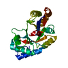
BIOMOLECULE: 1 THIS ENTRY CONTAINS THE CRYSTALLOGRAPHIC ASYMMETRIC UNIT WHICH CONSISTS OF 2 CHAIN(S) ...BIOMOLECULE: 1 THIS ENTRY CONTAINS THE CRYSTALLOGRAPHIC ASYMMETRIC UNIT WHICH CONSISTS OF 2 CHAIN(S). SEE REMARK 350 FOR INFORMATION ON GENERATING THE BIOLOGICAL MOLECULE(S). SIZE EXCLUSION CHROMATOGRAPHY WITH STATIC LIGHT SCATTERING SUPPORTS THE ASSIGNMENT OF THE DIMER AS A SIGNIFICANT OLIGOMERIZATION STATE.
Remark 999
SEQUENCE THE CONSTRUCT WAS EXPRESSED WITH A PURIFICATION TAG MGSDKIHHHHHHENLYFQG. THE TAG WAS ...SEQUENCE THE CONSTRUCT WAS EXPRESSED WITH A PURIFICATION TAG MGSDKIHHHHHHENLYFQG. THE TAG WAS REMOVED WITH TEV PROTEASE LEAVING ONLY A GLYCINE FOLLOWED BY THE TARGET SEQUENCE.
Resolution: 1.5→86.066 Å / Num. obs: 35176 / % possible obs: 76.7 % / Redundancy: 2.9 % / Rmerge(I) obs: 0.05 / Rsym value: 0.05 / Net I/σ(I): 9.4
Reflection shell
Diffraction-ID: 2
Resolution (Å)
Redundancy (%)
Rmerge(I) obs
Mean I/σ(I) obs
Num. measured all
Num. unique all
Rsym value
% possible all
1.5-1.54
2.7
0.183
4.1
5577
2100
0.183
61.3
1.54-1.58
2.7
0.153
4.8
5683
2129
0.153
64.9
1.58-1.63
2.7
0.134
5.5
5748
2126
0.134
65.5
1.63-1.68
2.7
0.112
6.2
5739
2113
0.112
67.4
1.68-1.73
2.8
0.105
7.2
5794
2085
0.105
70.4
1.73-1.79
2.8
0.093
8
5922
2115
0.093
72.6
1.79-1.86
2.8
0.079
9.4
6046
2122
0.079
74
1.86-1.94
3
0.07
10.4
6077
2052
0.07
75.3
1.94-2.02
3
0.063
11.4
6034
1985
0.063
75.5
2.02-2.12
3.2
0.056
12.8
6181
1958
0.056
79.8
2.12-2.24
3.2
0.057
12.2
6149
1944
0.057
81.9
2.24-2.37
3.1
0.059
11.7
5822
1876
0.059
83.7
2.37-2.54
3.1
0.056
12.3
5520
1799
0.056
85.8
2.54-2.74
3
0.048
14.1
5181
1715
0.048
86.5
2.74-3
3
0.044
15.7
4863
1616
0.044
89.4
3-3.35
3
0.046
14.1
4421
1480
0.046
92.1
3.35-3.87
2.9
0.042
15.6
3989
1357
0.042
92.7
3.87-4.74
3
0.037
16.7
3517
1174
0.037
97.3
4.74-6.71
3.2
0.041
16.5
2908
913
0.041
96.9
6.71-86.07
4.3
0.045
14.9
2237
517
0.045
99
-
Phasing
Phasing
Method: MAD
-
Processing
Software
Name
Version
Classification
NB
SHELX
refinement
MolProbity
3beta29
modelbuilding
SOLVE
phasing
SCALA
datascaling
PDB_EXTRACT
2
dataextraction
MAR345
CCD
datacollection
MOSFLM
datareduction
SHELXL-97
refinement
Refinement
Method to determine structure: MAD / Resolution: 1.5→28.736 Å / Num. parameters: 6902 / Num. restraintsaints: 9250 / Cross valid method: FREE R / Stereochemistry target values: ENGH AND HUBER Details: 1. HYDROGENS HAVE BEEN ADDED IN THE RIDING POSITIONS. 2. A MET-INHIBITION PROTOCOL WAS USED FOR SELENOMETHIONINE INCORPORATION DURING PROTEIN EXPRESSION. THE OCCUPANCY OF THE SE ATOMS IN THE ...Details: 1. HYDROGENS HAVE BEEN ADDED IN THE RIDING POSITIONS. 2. A MET-INHIBITION PROTOCOL WAS USED FOR SELENOMETHIONINE INCORPORATION DURING PROTEIN EXPRESSION. THE OCCUPANCY OF THE SE ATOMS IN THE MSE RESIDUES WAS REDUCED TO 0.75 TO ACCOUNT FOR THE REDUCED SCATTERING POWER DUE TO PARTIAL S-MET INCORPORATION. 3. DATA IS TETARTOHEDRALLY TWINNED. REFINEMENT WAS PERFORMED WITH THE TWIN OPERATORS (K,H,-L), (-K,-H,-L), AND (-H,-K,L) AND THE RESPECTIVE TWIN FRACTIONS OF 0.28, 0.19, 0.15. 4. THE NOMINAL RESOLUTION IS 1.65 A WITH 7312 OBSERVED REFLECTIONS BETWEEN 1.65-1.50 (64.2% COMPLETE FOR THIS SHELL) INCLUDED IN THE REFINEMENT. 5. THE R-FREE SET WAS GENERATED USING THE TWIN LAWS.
In the structure databanks used in Yorodumi, some data are registered as the other names, "COVID-19 virus" and "2019-nCoV". Here are the details of the virus and the list of structure data.
Jan 31, 2019. EMDB accession codes are about to change! (news from PDBe EMDB page)
EMDB accession codes are about to change! (news from PDBe EMDB page)
The allocation of 4 digits for EMDB accession codes will soon come to an end. Whilst these codes will remain in use, new EMDB accession codes will include an additional digit and will expand incrementally as the available range of codes is exhausted. The current 4-digit format prefixed with “EMD-” (i.e. EMD-XXXX) will advance to a 5-digit format (i.e. EMD-XXXXX), and so on. It is currently estimated that the 4-digit codes will be depleted around Spring 2019, at which point the 5-digit format will come into force.
The EM Navigator/Yorodumi systems omit the EMD- prefix.
Related info.:Q: What is EMD? / ID/Accession-code notation in Yorodumi/EM Navigator
Yorodumi is a browser for structure data from EMDB, PDB, SASBDB, etc.
This page is also the successor to EM Navigator detail page, and also detail information page/front-end page for Omokage search.
The word "yorodu" (or yorozu) is an old Japanese word meaning "ten thousand". "mi" (miru) is to see.
Related info.:EMDB / PDB / SASBDB / Comparison of 3 databanks / Yorodumi Search / Aug 31, 2016. New EM Navigator & Yorodumi / Yorodumi Papers / Jmol/JSmol / Function and homology information / Changes in new EM Navigator and Yorodumi
 Movie
Movie Controller

 Yorodumi
 Open data
 Basic information
 Components
 Keywords
 Function and homology information
 Shewanella loihica (bacteria)
 X-RAY DIFFRACTION /
  Authors
 Citation
 Structure visualization
 Downloads & links
 Other downloads
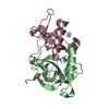


 PDBj
PDBj
 Assembly
 Mass: 18.015 Da / Num. of mol.: 9 / Source method: isolated from a natural source / Formula: H2O
 Sample preparation
 Processing