 Movie
Movie Controller

[English] 日本語
 Yorodumi
- PDB-2pr2: Structure of Mycobacterium tuberculosis enoyl-ACP reductase with ... -
+ Open data

- Basic information
| Entry | Database: PDB / ID: 2pr2 | ||||||
|---|---|---|---|---|---|---|---|
| Title | Structure of Mycobacterium tuberculosis enoyl-ACP reductase with bound INH-NADP. | ||||||
 Components Components | enoyl-ACP Reductase | ||||||
 Keywords Keywords | OXIDOREDUCTASE / drug target of isoniazid | ||||||
| Function / homology |  Function and homology information Function and homology informationenoyl-[acyl-carrier-protein] reductase [NAD(P)H] activity / trans-2-enoyl-CoA reductase (NADH) activity / mycolic acid biosynthetic process / enoyl-[acyl-carrier-protein] reductase (NADH) / fatty acid elongation / enoyl-[acyl-carrier-protein] reductase (NADH) activity / NAD+ binding / peptidoglycan-based cell wall / fatty acid binding / fatty acid biosynthetic process ...enoyl-[acyl-carrier-protein] reductase [NAD(P)H] activity / trans-2-enoyl-CoA reductase (NADH) activity / mycolic acid biosynthetic process / enoyl-[acyl-carrier-protein] reductase (NADH) / fatty acid elongation / enoyl-[acyl-carrier-protein] reductase (NADH) activity / NAD+ binding / peptidoglycan-based cell wall / fatty acid binding / fatty acid biosynthetic process / response to antibiotic / plasma membrane Similarity search - Function | ||||||
| Biological species |   Mycobacterium tuberculosis (bacteria) Mycobacterium tuberculosis (bacteria) | ||||||
| Method |  X-RAY DIFFRACTION / MOLECULAR REPLACEMENT / Resolution: 2.5 Å X-RAY DIFFRACTION / MOLECULAR REPLACEMENT / Resolution: 2.5 Å | ||||||
 Authors Authors | Vetting, M.W. / Argyrou, A. / Blanchard, J.S. | ||||||
 Citation Citation | Journal: J.Am.Chem.Soc. / Year: 2007 Title: New insight into the mechanism of action of and resistance to isoniazid: interaction of Mycobacterium tuberculosis enoyl-ACP reductase with INH-NADP Authors: Argyrou, A. / Vetting, M.W. / Blanchard, J.S. | ||||||
| History |
|
- Structure visualization
| Structure viewer | Molecule: MolmilJmol/JSmol |
|---|
- Downloads & links
-Download
| PDBx/mmCIF format | 2pr2.cif.gz | 66 KB | Display | PDBx/mmCIF format |
|---|---|---|---|---|
| PDB format | pdb2pr2.ent.gz | 47.8 KB | Display | PDB format |
| PDBx/mmJSON format | 2pr2.json.gz | Tree view | PDBx/mmJSON format | |
| Others |  Other downloads Other downloads |
-Validation report
| Arichive directory | https://data.pdbj.org/pub/pdb/validation_reports/pr/2pr2ftp://data.pdbj.org/pub/pdb/validation_reports/pr/2pr2 | HTTPS FTP |
|---|
-Related structure data
| Related structure data |  1enyS S: Starting model for refinement |
|---|---|
| Similar structure data |


-Links
 PDBj
PDBj

- Assembly
| Deposited unit | 
| ||||||||
|---|---|---|---|---|---|---|---|---|---|
| 1 | 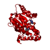
| ||||||||
| Unit cell |
| ||||||||
| Components on special symmetry positions |
| ||||||||
| Details | The biological assembly is monomeric and is contained within the assymetric unit. |
-Components
| #1: Protein | Mass: 28554.781 Da / Num. of mol.: 1 Source method: isolated from a genetically manipulated source Source: (gene. exp.) Mycobacterium tuberculosis (bacteria) / Strain: H37Rv / Gene: inhA / Plasmid: pET15b / Species (production host): Escherichia coli / Production host: References: UniProt: P0A5Y6, UniProt: P9WGR1*PLUS, enoyl-[acyl-carrier-protein] reductase (NADH) |
|---|---|
| #2: Chemical | ChemComp-DG1 / (  Mass: 850.515 Da / Num. of mol.: 1 / Source method: obtained synthetically / Formula: C27H33N8O18P3 Mass: 850.515 Da / Num. of mol.: 1 / Source method: obtained synthetically / Formula: C27H33N8O18P3 |
| #3: Water | ChemComp-HOH /  Mass: 18.015 Da / Num. of mol.: 42 / Source method: isolated from a natural source / Formula: H2O Mass: 18.015 Da / Num. of mol.: 42 / Source method: isolated from a natural source / Formula: H2O |
-Experimental details
-Experiment
| Experiment | Method: X-RAY DIFFRACTION / Number of used crystals: 1 |
|---|
- Sample preparation
| Crystal | Density Matthews: 3.46 Å3/Da / Density % sol: 64.42 % |
|---|---|
| Crystal grow | Temperature: 291 K / Method: vapor diffusion / pH: 9 Details: Cocrystallization with Inh-NADP at 4.8 mM 20 25% of 2-methyl-2,4-pentanediol, 100 mM Bicine pH 9.0., Vapour diffusion under oil, temperature 291K, VAPOR DIFFUSION |
-Data collection
| Diffraction | Mean temperature: 193 K |
|---|---|
| Diffraction source | Source: ROTATING ANODE / Type: RIGAKU RU300 / Wavelength: 1.5418 Å |
| Detector | Type: RIGAKU RAXIS IV++ / Detector: IMAGE PLATE / Date: Dec 12, 2005 |
| Radiation | Monochromator: Rigaku Osmic Blue optic / Protocol: SINGLE WAVELENGTH / Monochromatic (M) / Laue (L): M / Scattering type: x-ray |
| Radiation wavelength | Wavelength: 1.5418 Å / Relative weight: 1 |
| Reflection | Resolution: 2.5→41 Å / Num. all: 14562 / Num. obs: 14562 / % possible obs: 99.8 % / Observed criterion σ(F): 0 / Observed criterion σ(I): 0 / Redundancy: 11.6 % / Biso Wilson estimate: 51.9 Å2 / Rmerge(I) obs: 0.053 / Rsym value: 0.055 / Net I/σ(I): 36.7 |
| Reflection shell | Resolution: 2.5→2.64 Å / Redundancy: 7.5 % / Rmerge(I) obs: 0.337 / Mean I/σ(I) obs: 7.5 / Num. unique all: 2077 / Rsym value: 0.353 / % possible all: 100 |
- Processing
| Software |
| ||||||||||||||||||||||||||||||||||||||||||||||||||||||||||||||||||||||||||||||||||||||||||
|---|---|---|---|---|---|---|---|---|---|---|---|---|---|---|---|---|---|---|---|---|---|---|---|---|---|---|---|---|---|---|---|---|---|---|---|---|---|---|---|---|---|---|---|---|---|---|---|---|---|---|---|---|---|---|---|---|---|---|---|---|---|---|---|---|---|---|---|---|---|---|---|---|---|---|---|---|---|---|---|---|---|---|---|---|---|---|---|---|---|---|---|
| Refinement | Method to determine structure: MOLECULAR REPLACEMENT Starting model: PDB ENTRY 1ENY Resolution: 2.5→41 Å / Cor.coef. Fo:Fc: 0.953 / Cor.coef. Fo:Fc free: 0.939 / SU B: 12.636 / SU ML: 0.145 / TLS residual ADP flag: LIKELY RESIDUAL / Cross valid method: THROUGHOUT / σ(F): 0 / σ(I): 0 / ESU R: 0.289 / ESU R Free: 0.215 / Stereochemistry target values: MAXIMUM LIKELIHOOD / Details: HYDROGENS HAVE BEEN ADDED IN THE RIDING POSITIONS
| ||||||||||||||||||||||||||||||||||||||||||||||||||||||||||||||||||||||||||||||||||||||||||
| Solvent computation | Ion probe radii: 0.8 Å / Shrinkage radii: 0.8 Å / VDW probe radii: 1.2 Å / Solvent model: MASK | ||||||||||||||||||||||||||||||||||||||||||||||||||||||||||||||||||||||||||||||||||||||||||
| Displacement parameters | Biso mean: 37.888 Å2
| ||||||||||||||||||||||||||||||||||||||||||||||||||||||||||||||||||||||||||||||||||||||||||
| Refinement step | Cycle: LAST / Resolution: 2.5→41 Å
| ||||||||||||||||||||||||||||||||||||||||||||||||||||||||||||||||||||||||||||||||||||||||||
| Refine LS restraints |
| ||||||||||||||||||||||||||||||||||||||||||||||||||||||||||||||||||||||||||||||||||||||||||
| LS refinement shell | Resolution: 2.5→2.565 Å / Total num. of bins used: 20
| ||||||||||||||||||||||||||||||||||||||||||||||||||||||||||||||||||||||||||||||||||||||||||
| Refinement TLS params. | Method: refined / Origin x: 3.648 Å / Origin y: -37.408 Å / Origin z: 18.167 Å
|
