 Movie
Movie Controller


+ Open data

- Basic information
| Entry | Database: PDB / ID: 2pnr | ||||||
|---|---|---|---|---|---|---|---|
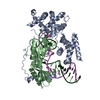
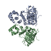
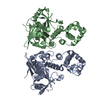
| Title | Crystal Structure of the asymmetric Pdk3-l2 Complex | ||||||
 Components Components |
| ||||||
 Keywords Keywords | TRANSFERASE / pyruvate dehydrogenase kinase 3 / lipoyl-bearing domain / asymmetric protein-protein complex | ||||||
| Function / homology |  Function and homology information Function and homology informationhypoxia-inducible factor-1alpha signaling pathway / PDH complex synthesizes acetyl-CoA from PYR / [pyruvate dehydrogenase (acetyl-transferring)] kinase / pyruvate dehydrogenase (acetyl-transferring) kinase activity / regulation of pyruvate decarboxylation to acetyl-CoA / dihydrolipoyllysine-residue acetyltransferase / dihydrolipoyllysine-residue acetyltransferase activity / pyruvate decarboxylation to acetyl-CoA / Regulation of pyruvate dehydrogenase (PDH) complex / Protein lipoylation ...hypoxia-inducible factor-1alpha signaling pathway / PDH complex synthesizes acetyl-CoA from PYR / [pyruvate dehydrogenase (acetyl-transferring)] kinase / pyruvate dehydrogenase (acetyl-transferring) kinase activity / regulation of pyruvate decarboxylation to acetyl-CoA / dihydrolipoyllysine-residue acetyltransferase / dihydrolipoyllysine-residue acetyltransferase activity / pyruvate decarboxylation to acetyl-CoA / Regulation of pyruvate dehydrogenase (PDH) complex / Protein lipoylation / pyruvate catabolic process / pyruvate dehydrogenase complex / cellular response to fatty acid / regulation of reactive oxygen species metabolic process / Signaling by Retinoic Acid / regulation of glucose metabolic process / tricarboxylic acid cycle / peroxisome proliferator activated receptor signaling pathway / peptidyl-serine phosphorylation / cellular response to glucose stimulus / glucose metabolic process / protein kinase activity / mitochondrial matrix / protein serine/threonine kinase activity / nucleolus / mitochondrion / ATP binding / identical protein binding Similarity search - Function | ||||||
| Biological species |  Homo sapiens (human) Homo sapiens (human) | ||||||
| Method |  X-RAY DIFFRACTION / SYNCHROTRON / MOLECULAR REPLACEMENT / Resolution: 2.5 Å X-RAY DIFFRACTION / SYNCHROTRON / MOLECULAR REPLACEMENT / Resolution: 2.5 Å | ||||||
 Authors Authors | Vassylyev, D.G. / Steussy, C.N. / Devedjiev, Y. | ||||||
 Citation Citation | Journal: J.Mol.Biol. / Year: 2007 Title: Crystal Structure of an Asymmetric complex of Pyruvate Dehydrogenase Kinase 3 with Lipoyl domain 2 and its Biological Implications Authors: Devedjiev, Y. / Steussy, C.N. / Vassylyev, D.G. | ||||||
| History |
|
- Structure visualization
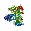
| Structure viewer | Molecule: MolmilJmol/JSmol |
|---|
- Downloads & links
-Download
| PDBx/mmCIF format | 2pnr.cif.gz | 342.3 KB | Display | PDBx/mmCIF format |
|---|---|---|---|---|
| PDB format | pdb2pnr.ent.gz | 275.8 KB | Display | PDB format |
| PDBx/mmJSON format | 2pnr.json.gz | Tree view | PDBx/mmJSON format | |
| Others |  Other downloads Other downloads |
-Validation report
| Arichive directory | https://data.pdbj.org/pub/pdb/validation_reports/pn/2pnrftp://data.pdbj.org/pub/pdb/validation_reports/pn/2pnr | HTTPS FTP |
|---|
-Related structure data
| Related structure data | 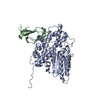 1y8nS S: Starting model for refinement |
|---|---|
| Similar structure data |


-Links
 PDBj
PDBj


- Assembly
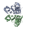
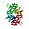
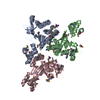
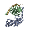
| Deposited unit | 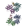
| ||||||||
|---|---|---|---|---|---|---|---|---|---|
| 1 | 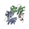
| ||||||||
| 2 | 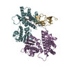
| ||||||||
| Unit cell |
|
-Components
| #1: Protein | [ Mass: 48290.980 Da / Num. of mol.: 4 Source method: isolated from a genetically manipulated source Source: (gene. exp.) Homo sapiens (human) / Gene: PDK3 / Plasmid: PTRCHISB / Species (production host): Escherichia coli / Production host:  References: UniProt: Q15120, [pyruvate dehydrogenase (acetyl-transferring)] kinase #2: Protein | Mass: 14192.097 Da / Num. of mol.: 2 Source method: isolated from a genetically manipulated source Source: (gene. exp.) Homo sapiens (human) / Gene: DLAT, DLTA / Plasmid: PTRCHISB / Species (production host): Escherichia coli / Production host: References: UniProt: P10515, dihydrolipoyllysine-residue acetyltransferase #3: Chemical | 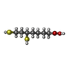  Mass: 208.341 Da / Num. of mol.: 2 / Source method: obtained synthetically / Formula: C8H16O2S2 Mass: 208.341 Da / Num. of mol.: 2 / Source method: obtained synthetically / Formula: C8H16O2S2#4: Water | ChemComp-HOH / |  Mass: 18.015 Da / Num. of mol.: 723 / Source method: isolated from a natural source / Formula: H2O Mass: 18.015 Da / Num. of mol.: 723 / Source method: isolated from a natural source / Formula: H2OHas protein modification | Y | |
|---|
-Experimental details
-Experiment
| Experiment | Method: X-RAY DIFFRACTION |
|---|
- Sample preparation
| Crystal | Density Matthews: 2.32 Å3/Da / Density % sol: 47.1 % |
|---|---|
| Crystal grow | Temperature: 295 K / Method: vapor diffusion, sitting drop / pH: 8 Details: Protein solution: 5mg/ml, 20mM Tris-HCL pH 8.0, 100mM NaCl, 5mM DTT, 2.5% (v/v) ethylen glycol Precipitant solution: 100mM histidine, 30 mM EDTA, 7% (v/v) MPD., VAPOR DIFFUSION, SITTING DROP, temperature 295K |
-Data collection
| Diffraction | Mean temperature: 100 K |
|---|---|
| Diffraction source | Source: SYNCHROTRON / Site: APS  / Beamline: 17-ID / Wavelength: 1 Å / Beamline: 17-ID / Wavelength: 1 Å |
| Detector | Type: MAR CCD 165 mm / Detector: CCD / Date: Mar 18, 2002 |
| Radiation | Monochromator: Graphite / Protocol: SINGLE WAVELENGTH / Monochromatic (M) / Laue (L): M / Scattering type: x-ray |
| Radiation wavelength | Wavelength: 1 Å / Relative weight: 1 |
| Reflection | Resolution: 2.5→30 Å / Num. all: 64645 / Num. obs: 64645 / % possible obs: 92.9 % / Observed criterion σ(F): 0 / Observed criterion σ(I): 0 / Redundancy: 4.3 % / Biso Wilson estimate: 41 Å2 / Rmerge(I) obs: 0.097 / Rsym value: 0.097 / Net I/σ(I): 12.1 |
| Reflection shell | Resolution: 2.5→2.59 Å / Redundancy: 1.9 % / Rmerge(I) obs: 0.422 / Mean I/σ(I) obs: 1.5 / Num. unique all: 4439 / Rsym value: 0.422 / % possible all: 64 |
- Processing
| Software |
| |||||||||||||||||||||||||
|---|---|---|---|---|---|---|---|---|---|---|---|---|---|---|---|---|---|---|---|---|---|---|---|---|---|---|
| Refinement | Method to determine structure: MOLECULAR REPLACEMENT Starting model: 1Y8N Resolution: 2.5→30 Å / Isotropic thermal model: isotropic / Cross valid method: THROUGHOUT / σ(F): 0 / σ(I): 0 / Stereochemistry target values: Engh & Huber Details: THE PERFECT MEROHEDRAL TWINNING WAS DETECTED IN THE CRYSTALS WITH THE TWINNING OPERATOR {H,-K,-L}. THE REFINEMENT STATISTICS PRESENTED FOR THIS ENTRY CORRESPONDS TO THE REFINEMENT CARRIED ...Details: THE PERFECT MEROHEDRAL TWINNING WAS DETECTED IN THE CRYSTALS WITH THE TWINNING OPERATOR {H,-K,-L}. THE REFINEMENT STATISTICS PRESENTED FOR THIS ENTRY CORRESPONDS TO THE REFINEMENT CARRIED OUT USING THE TWINNING OPTION OF THE CNS PROGRAM. FOR THE DEPOSITION THE DIFFRACTION DATA WERE DETWINNED USING THE CNS PROGRAM. THE REFINEMENT STATISTICS CALCULATED BASED ON THE DETWINNED DATA MIGHT BE SLIGHTLY DIFFERENT FROM THOSE OBTAINED DURING THE "TWINNED" REFINEMENT INCLUDED IN THIS ENTRY.
| |||||||||||||||||||||||||
| Refine analyze |
| |||||||||||||||||||||||||
| Refinement step | Cycle: LAST / Resolution: 2.5→30 Å
| |||||||||||||||||||||||||
| Refine LS restraints |
| |||||||||||||||||||||||||
| LS refinement shell | Resolution: 2.5→2.59 Å
|
