 Movie
Movie Controller

[English] 日本語
 Yorodumi
- PDB-2pe5: Crystal Structure of the Lac Repressor bound to ONPG in repressed... -
+ Open data

- Basic information
| Entry | Database: PDB / ID: 2pe5 | |||||||||
|---|---|---|---|---|---|---|---|---|---|---|
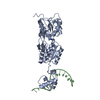
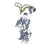
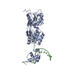
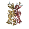
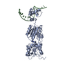
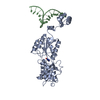
| Title | Crystal Structure of the Lac Repressor bound to ONPG in repressed state | |||||||||
 Components Components |
| |||||||||
 Keywords Keywords | TRANSCRIPTION/DNA / Lac repressor / allosteric effectors / gene regulation / DNA-binding / Transcription / Transcription regulation / TRANSCRIPTION-DNA COMPLEX | |||||||||
| Function / homology |  Function and homology information Function and homology informationDNA-binding transcription repressor activity / cis-regulatory region sequence-specific DNA binding / transcription cis-regulatory region binding / DNA-binding transcription factor activity / negative regulation of DNA-templated transcription / regulation of DNA-templated transcription / identical protein binding / cytosol Similarity search - Function | |||||||||
| Biological species |  synthetic construct (others) | |||||||||
| Method |  X-RAY DIFFRACTION / MOLECULAR REPLACEMENT / Resolution: 3.5 Å X-RAY DIFFRACTION / MOLECULAR REPLACEMENT / Resolution: 3.5 Å | |||||||||
 Authors Authors | Daber, R. / Stayrook, S.E. / Rosenberg, A. / Lewis, M. | |||||||||
 Citation Citation | Journal: J.Mol.Biol. / Year: 2007 Title: Structural analysis of lac repressor bound to allosteric effectors Authors: Daber, R. / Stayrook, S. / Rosenberg, A. / Lewis, M. | |||||||||
| History |
|
- Structure visualization
| Structure viewer | Molecule: MolmilJmol/JSmol |
|---|
- Downloads & links
-Download
| PDBx/mmCIF format | 2pe5.cif.gz | 222.3 KB | Display | PDBx/mmCIF format |
|---|---|---|---|---|
| PDB format | pdb2pe5.ent.gz | 174.9 KB | Display | PDB format |
| PDBx/mmJSON format | 2pe5.json.gz | Tree view | PDBx/mmJSON format | |
| Others |  Other downloads Other downloads |
-Validation report
| Arichive directory | https://data.pdbj.org/pub/pdb/validation_reports/pe/2pe5ftp://data.pdbj.org/pub/pdb/validation_reports/pe/2pe5 | HTTPS FTP |
|---|
-Related structure data
| Related structure data |  2p9hC  2pafC  1efaS C: citing same article ( S: Starting model for refinement |
|---|---|
| Similar structure data |


-Links
 PDBj
PDBj


- Assembly
| Deposited unit | 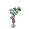
| ||||||||
|---|---|---|---|---|---|---|---|---|---|
| 1 | 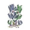
| ||||||||
| 2 | 
| ||||||||
| Unit cell |
|
-Components
| #1: DNA chain | Mass: 6132.991 Da / Num. of mol.: 3 / Source method: obtained synthetically / Source: (synth.) synthetic construct (others) #2: Protein | Mass: 35410.438 Da / Num. of mol.: 3 / Fragment: sequence database residues 2-331 / Mutation: S61L Source method: isolated from a genetically manipulated source Source: (gene. exp.) #3: Sugar |   Type: D-saccharide / Mass: 301.249 Da / Num. of mol.: 3 / Source method: obtained synthetically / Formula: C12H15NO8 Type: D-saccharide / Mass: 301.249 Da / Num. of mol.: 3 / Source method: obtained synthetically / Formula: C12H15NO8 |
|---|
-Experimental details
-Experiment
| Experiment | Method: X-RAY DIFFRACTION / Number of used crystals: 1 |
|---|
- Sample preparation
| Crystal | Density Matthews: 5.03 Å3/Da / Density % sol: 75.52 % | ||||||||||||||||||||||||||||||||||||||||||||
|---|---|---|---|---|---|---|---|---|---|---|---|---|---|---|---|---|---|---|---|---|---|---|---|---|---|---|---|---|---|---|---|---|---|---|---|---|---|---|---|---|---|---|---|---|---|
| Crystal grow | Temperature: 281 K / Method: vapor diffusion, hanging drop / pH: 7.5 Details: 1.8 M Ammonium Sulfate, 0.1 M Hepes pH 7.5. 11% PEG 400, 14% glycerol, 10X ONPG, VAPOR DIFFUSION, HANGING DROP, temperature 281K | ||||||||||||||||||||||||||||||||||||||||||||
| Components of the solutions |
|
-Data collection
| Diffraction | Mean temperature: 100 K |
|---|---|
| Diffraction source | Source: ROTATING ANODE / Type: RIGAKU / Wavelength: 1.5418 Å |
| Detector | Type: MAR scanner 300 mm plate / Detector: IMAGE PLATE / Date: Mar 19, 2006 / Details: Osmic mirrors |
| Radiation | Monochromator: Osmic mirrors / Protocol: SINGLE WAVELENGTH / Monochromatic (M) / Laue (L): M / Scattering type: x-ray |
| Radiation wavelength | Wavelength: 1.5418 Å / Relative weight: 1 |
| Reflection | Resolution: 3.5→20 Å / Num. all: 31342 / Num. obs: 29717 / % possible obs: 99.89 % / Observed criterion σ(F): -3 / Observed criterion σ(I): -3 / Redundancy: 9.1 % / Biso Wilson estimate: 25.4 Å2 / Rmerge(I) obs: 0.176 / Rsym value: 0.176 / Net I/σ(I): 7.3 |
| Reflection shell | Resolution: 3.5→3.6 Å / Redundancy: 6.9 % / Rmerge(I) obs: 0.504 / Mean I/σ(I) obs: 4.37 / Num. unique all: 2090 / Rsym value: 0.504 / % possible all: 99.7 |
- Processing
| Software |
| ||||||||||||||||||||||||||||||||||||||||||||||||||||||||||||||||||||||||||||||||||||||||||
|---|---|---|---|---|---|---|---|---|---|---|---|---|---|---|---|---|---|---|---|---|---|---|---|---|---|---|---|---|---|---|---|---|---|---|---|---|---|---|---|---|---|---|---|---|---|---|---|---|---|---|---|---|---|---|---|---|---|---|---|---|---|---|---|---|---|---|---|---|---|---|---|---|---|---|---|---|---|---|---|---|---|---|---|---|---|---|---|---|---|---|---|
| Refinement | Method to determine structure: MOLECULAR REPLACEMENT Starting model: PDB entry 1EFA Resolution: 3.5→19.98 Å / Cor.coef. Fo:Fc: 0.878 / Cor.coef. Fo:Fc free: 0.82 / SU B: 24.027 / SU ML: 0.398 / Cross valid method: THROUGHOUT / σ(F): 0 / σ(I): 0 / ESU R Free: 0.548 / Stereochemistry target values: MAXIMUM LIKELIHOOD / Details: HYDROGENS HAVE BEEN ADDED IN THE RIDING POSITIONS
| ||||||||||||||||||||||||||||||||||||||||||||||||||||||||||||||||||||||||||||||||||||||||||
| Solvent computation | Ion probe radii: 0.8 Å / Shrinkage radii: 0.8 Å / VDW probe radii: 1.2 Å / Solvent model: MASK | ||||||||||||||||||||||||||||||||||||||||||||||||||||||||||||||||||||||||||||||||||||||||||
| Displacement parameters | Biso mean: 53.844 Å2
| ||||||||||||||||||||||||||||||||||||||||||||||||||||||||||||||||||||||||||||||||||||||||||
| Refinement step | Cycle: LAST / Resolution: 3.5→19.98 Å
| ||||||||||||||||||||||||||||||||||||||||||||||||||||||||||||||||||||||||||||||||||||||||||
| Refine LS restraints |
| ||||||||||||||||||||||||||||||||||||||||||||||||||||||||||||||||||||||||||||||||||||||||||
| LS refinement shell | Resolution: 3.5→3.589 Å / Total num. of bins used: 20
|
