- PDB-2pbz: Crystal structure of an IMP biosynthesis protein PurP from Thermo... -
+
Open data
ID or keywords:
Loading...
-
Basic information
Entry
Database: PDB / ID: 2pbz
Title
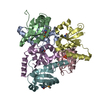
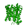
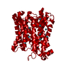
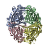
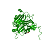
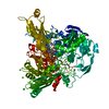
Crystal structure of an IMP biosynthesis protein PurP from Thermococcus kodakaraensis
Components
Hypothetical protein
Keywords
LIGASE / NYSGXRC / PSI-II / IMP biosynthesis / ATP binding protein / PurP / 10188d / Structural Genomics / Protein Structure Initiative / New York SGX Research Center for Structural Genomics
Function / homology
Function and homology information
IMP biosynthetic process / ligase activity, forming carbon-nitrogen bonds / magnesium ion binding / ATP binding Similarity search - Function
IMP biosynthesis enzyme PurP, C-terminal / IMP biosynthesis enzyme PurP, N-terminal / IMP biosynthesis enzyme PurP / Protein of unknown function (DUF1246) / Domain of unknown function (DUF1297) / Rossmann fold - #20 / ATP-grasp fold, A domain / ATP-grasp fold, subdomain 1 / ATP-grasp fold, B domain / Pre-ATP-grasp domain superfamily ...IMP biosynthesis enzyme PurP, C-terminal / IMP biosynthesis enzyme PurP, N-terminal / IMP biosynthesis enzyme PurP / Protein of unknown function (DUF1246) / Domain of unknown function (DUF1297) / Rossmann fold - #20 / ATP-grasp fold, A domain / ATP-grasp fold, subdomain 1 / ATP-grasp fold, B domain / Pre-ATP-grasp domain superfamily / D-amino Acid Aminotransferase; Chain A, domain 1 / Dna Ligase; domain 1 / Rossmann fold / 2-Layer Sandwich / 3-Layer(aba) Sandwich / Alpha Beta Similarity search - Domain/homology
Monochromator: SI(III) / Protocol: SINGLE WAVELENGTH / Monochromatic (M) / Laue (L): M / Scattering type: x-ray
Radiation wavelength
Wavelength: 0.9792 Å / Relative weight: 1
Reflection
Resolution: 2.5→50 Å / Num. obs: 33174 / % possible obs: 94.7 % / Observed criterion σ(I): 0 / Redundancy: 12 % / Biso Wilson estimate: 25.5 Å2 / Rmerge(I) obs: 0.05 / Net I/σ(I): 12.5
Reflection shell
Resolution: 2.5→2.59 Å / Redundancy: 5.4 % / Rmerge(I) obs: 0.3 / Mean I/σ(I) obs: 1 / % possible all: 68.1
-
Processing
Software
Name
Version
Classification
CNS
1.1
refinement
HKL-2000
datareduction
HKL-2000
datascaling
SHARP
phasing
SHELXD
phasing
Refinement
Method to determine structure: SAD / Resolution: 2.5→31 Å / Rfactor Rfree error: 0.011 / Data cutoff high absF: 90726.93 / Data cutoff low absF: 0 / Isotropic thermal model: RESTRAINED / Cross valid method: THROUGHOUT / σ(F): 2 / Stereochemistry target values: ENGH & HUBER Details: 1. Restrained NCS was used during refinement due to paucity of data. 2. Residual density in the final difference map was modeled as ATP molecule. 3. Residues listed as missing in remark 465 ...Details: 1. Restrained NCS was used during refinement due to paucity of data. 2. Residual density in the final difference map was modeled as ATP molecule. 3. Residues listed as missing in remark 465 were not modeled because of lack of electron density. 4. Outliers for Ramachandran plot were due to poor electron density except for His11 of all chains. The electron density for His 11 is well defined.
In the structure databanks used in Yorodumi, some data are registered as the other names, "COVID-19 virus" and "2019-nCoV". Here are the details of the virus and the list of structure data.
Jan 31, 2019. EMDB accession codes are about to change! (news from PDBe EMDB page)
EMDB accession codes are about to change! (news from PDBe EMDB page)
The allocation of 4 digits for EMDB accession codes will soon come to an end. Whilst these codes will remain in use, new EMDB accession codes will include an additional digit and will expand incrementally as the available range of codes is exhausted. The current 4-digit format prefixed with “EMD-” (i.e. EMD-XXXX) will advance to a 5-digit format (i.e. EMD-XXXXX), and so on. It is currently estimated that the 4-digit codes will be depleted around Spring 2019, at which point the 5-digit format will come into force.
The EM Navigator/Yorodumi systems omit the EMD- prefix.
Related info.:Q: What is EMD? / ID/Accession-code notation in Yorodumi/EM Navigator
Yorodumi is a browser for structure data from EMDB, PDB, SASBDB, etc.
This page is also the successor to EM Navigator detail page, and also detail information page/front-end page for Omokage search.
The word "yorodu" (or yorozu) is an old Japanese word meaning "ten thousand". "mi" (miru) is to see.
Related info.:EMDB / PDB / SASBDB / Comparison of 3 databanks / Yorodumi Search / Aug 31, 2016. New EM Navigator & Yorodumi / Yorodumi Papers / Jmol/JSmol / Function and homology information / Changes in new EM Navigator and Yorodumi
 Movie
Movie Controller

 Yorodumi
 Open data
 Basic information
 Components
 Keywords
 Function and homology information
 Thermococcus kodakarensis (archaea)
 X-RAY DIFFRACTION /
  Authors
 Citation
 Structure visualization
 Downloads & links
 Other downloads
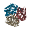

 PDBj
PDBj


 Assembly
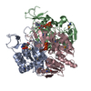


 Mass: 507.181 Da / Num. of mol.: 3 / Source method: obtained synthetically / Formula: C10H16N5O13P3 / Comment: ATP, energy-carrying molecule*YM
 Mass: 18.015 Da / Num. of mol.: 240 / Source method: isolated from a natural source / Formula: H2O
 Sample preparation
 Processing