 Movie
Movie Controller

[English] 日本語
 Yorodumi
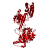
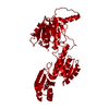
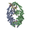
- PDB-2p9g: Crystal structure of serine bound G336V,G337V double mutant of E.... -
+ Open data

- Basic information
| Entry | Database: PDB / ID: 2p9g | ||||||
|---|---|---|---|---|---|---|---|
| Title | Crystal structure of serine bound G336V,G337V double mutant of E.coli phosphoglycerate dehydrogenase | ||||||
 Components Components | D-3-phosphoglycerate dehydrogenase | ||||||
 Keywords Keywords | OXIDOREDUCTASE / serine biosynthesis / D-3-phosphoglycerate dehydrogenase / double mutation | ||||||
| Function / homology |  Function and homology information Function and homology information(S)-2-hydroxyglutarate dehydrogenase activity / 2-oxoglutarate reductase / phosphoglycerate dehydrogenase / phosphoglycerate dehydrogenase activity / serine binding / NADH binding / L-serine biosynthetic process / NAD+ binding / identical protein binding / cytosol Similarity search - Function | ||||||
| Biological species |  | ||||||
| Method |  X-RAY DIFFRACTION / MOLECULAR REPLACEMENT / Resolution: 2.8 Å X-RAY DIFFRACTION / MOLECULAR REPLACEMENT / Resolution: 2.8 Å | ||||||
 Authors Authors | Dey, S. / Sacchettini, J.C. | ||||||
 Citation Citation | Journal: J.Biol.Chem. / Year: 2007 Title: The Effect of Hinge Mutations on Effector Binding and Domain Rotation in Escherichia coli D-3-Phosphoglycerate Dehydrogenase. Authors: Dey, S. / Hu, Z. / Xu, X.L. / Sacchettini, J.C. / Grant, G.A. | ||||||
| History |
|
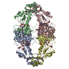
- Structure visualization
| Structure viewer | Molecule: MolmilJmol/JSmol |
|---|
- Downloads & links
-Download
| PDBx/mmCIF format | 2p9g.cif.gz | 165.5 KB | Display | PDBx/mmCIF format |
|---|---|---|---|---|
| PDB format | pdb2p9g.ent.gz | 131.3 KB | Display | PDB format |
| PDBx/mmJSON format | 2p9g.json.gz | Tree view | PDBx/mmJSON format | |
| Others |  Other downloads Other downloads |
-Validation report
| Summary document | 2p9g_validation.pdf.gz | 964.2 KB | Display | wwPDB validaton report |
|---|---|---|---|---|
| Full document | 2p9g_full_validation.pdf.gz | 995.1 KB | Display | |
| Data in XML | 2p9g_validation.xml.gz | 38.2 KB | Display | |
| Data in CIF | 2p9g_validation.cif.gz | 49.1 KB | Display | |
| Arichive directory | https://data.pdbj.org/pub/pdb/validation_reports/p9/2p9gftp://data.pdbj.org/pub/pdb/validation_reports/p9/2p9g | HTTPS FTP |
-Related structure data
| Related structure data | 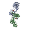 2p9cC 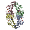 2p9eC  2pa3C 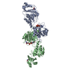 1psdS C: citing same article ( S: Starting model for refinement |
|---|---|
| Similar structure data |


-Links
 PDBj
PDBj


- Assembly
| Deposited unit | 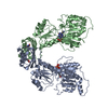
| ||||||||
|---|---|---|---|---|---|---|---|---|---|
| 1 | 
| ||||||||
| Unit cell |
|
-Components
| #1: Protein | Mass: 44182.527 Da / Num. of mol.: 2 / Mutation: C81A, C83A, C250A, C282A, G336V, G337V Source method: isolated from a genetically manipulated source Source: (gene. exp.) #2: Chemical |   Mass: 665.441 Da / Num. of mol.: 2 / Source method: obtained synthetically / Formula: C21H29N7O14P2 Mass: 665.441 Da / Num. of mol.: 2 / Source method: obtained synthetically / Formula: C21H29N7O14P2#3: Chemical |   Type: L-peptide linking / Mass: 105.093 Da / Num. of mol.: 2 / Source method: obtained synthetically / Formula: C3H7NO3 Type: L-peptide linking / Mass: 105.093 Da / Num. of mol.: 2 / Source method: obtained synthetically / Formula: C3H7NO3#4: Water | ChemComp-HOH / |  Mass: 18.015 Da / Num. of mol.: 49 / Source method: isolated from a natural source / Formula: H2O Mass: 18.015 Da / Num. of mol.: 49 / Source method: isolated from a natural source / Formula: H2O |
|---|
-Experimental details
-Experiment
| Experiment | Method: X-RAY DIFFRACTION / Number of used crystals: 1 |
|---|
- Sample preparation
| Crystal | Density Matthews: 2.82 Å3/Da / Density % sol: 56.41 % |
|---|---|
| Crystal grow | Temperature: 291 K / Method: vapor diffusion, hanging drop / pH: 4.5 Details: 30% PEG 400, 0.1M acetate, 0.2M Calcium acetate, pH 4.5, VAPOR DIFFUSION, HANGING DROP, temperature 291K |
-Data collection
| Diffraction source | Source: ROTATING ANODE / Type: OTHER / Wavelength: 1.541 Å |
|---|---|
| Detector | Type: RIGAKU RAXIS IV / Detector: IMAGE PLATE / Date: May 9, 2006 / Details: monochromator |
| Radiation | Protocol: SINGLE WAVELENGTH / Monochromatic (M) / Laue (L): M / Scattering type: x-ray |
| Radiation wavelength | Wavelength: 1.541 Å / Relative weight: 1 |
| Reflection | Resolution: 2.8→40.32 Å / Num. obs: 25289 / % possible obs: 99.5 % / Redundancy: 3.3 % / Rmerge(I) obs: 0.052 / Net I/σ(I): 20.1 |
| Reflection shell | Resolution: 2.8→2.95 Å / Redundancy: 3.3 % / Rmerge(I) obs: 0.207 / Mean I/σ(I) obs: 4.8 / % possible all: 100 |
- Processing
| Software |
| |||||||||||||||||||||||||||||||||||||||||||||||||||||||||||||||||||||||||||||||||||||
|---|---|---|---|---|---|---|---|---|---|---|---|---|---|---|---|---|---|---|---|---|---|---|---|---|---|---|---|---|---|---|---|---|---|---|---|---|---|---|---|---|---|---|---|---|---|---|---|---|---|---|---|---|---|---|---|---|---|---|---|---|---|---|---|---|---|---|---|---|---|---|---|---|---|---|---|---|---|---|---|---|---|---|---|---|---|---|
| Refinement | Method to determine structure: MOLECULAR REPLACEMENT Starting model: pdb entry 1PSD Resolution: 2.8→35 Å / Cor.coef. Fo:Fc: 0.936 / Cor.coef. Fo:Fc free: 0.911 / SU B: 13.366 / SU ML: 0.269 / Cross valid method: THROUGHOUT / ESU R Free: 0.366 / Stereochemistry target values: MAXIMUM LIKELIHOOD
| |||||||||||||||||||||||||||||||||||||||||||||||||||||||||||||||||||||||||||||||||||||
| Solvent computation | Ion probe radii: 0.8 Å / Shrinkage radii: 0.8 Å / VDW probe radii: 1.4 Å / Solvent model: MASK | |||||||||||||||||||||||||||||||||||||||||||||||||||||||||||||||||||||||||||||||||||||
| Displacement parameters | Biso mean: 44.721 Å2
| |||||||||||||||||||||||||||||||||||||||||||||||||||||||||||||||||||||||||||||||||||||
| Refinement step | Cycle: LAST / Resolution: 2.8→35 Å
| |||||||||||||||||||||||||||||||||||||||||||||||||||||||||||||||||||||||||||||||||||||
| Refine LS restraints |
| |||||||||||||||||||||||||||||||||||||||||||||||||||||||||||||||||||||||||||||||||||||
| LS refinement shell | Resolution: 2.8→2.872 Å / Total num. of bins used: 20
|
