 Movie
Movie Controller

[English] 日本語
 Yorodumi
- PDB-2p9e: Crystal Structure of G336V mutant of E.coli phosphoglycerate dehy... -
+ Open data

- Basic information
| Entry | Database: PDB / ID: 2p9e | ||||||
|---|---|---|---|---|---|---|---|
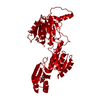
| Title | Crystal Structure of G336V mutant of E.coli phosphoglycerate dehydrogenase | ||||||
 Components Components | D-3-phosphoglycerate dehydrogenase | ||||||
 Keywords Keywords | OXIDOREDUCTASE / G336V mutant / phosphoglycerate dehydrogenase / serine biosynthesis | ||||||
| Function / homology |  Function and homology information Function and homology information(S)-2-hydroxyglutarate dehydrogenase activity / 2-oxoglutarate reductase / phosphoglycerate dehydrogenase / phosphoglycerate dehydrogenase activity / serine binding / NADH binding / L-serine biosynthetic process / NAD+ binding / identical protein binding / cytosol Similarity search - Function | ||||||
| Biological species |  | ||||||
| Method |  X-RAY DIFFRACTION / SYNCHROTRON / MOLECULAR REPLACEMENT / Resolution: 2.6 Å X-RAY DIFFRACTION / SYNCHROTRON / MOLECULAR REPLACEMENT / Resolution: 2.6 Å | ||||||
 Authors Authors | Dey, S. / Sacchettini, J.C. | ||||||
 Citation Citation | Journal: J.Biol.Chem. / Year: 2007 Title: The Effect of Hinge Mutations on Effector Binding and Domain Rotation in Escherichia coli D-3-Phosphoglycerate Dehydrogenase Authors: Dey, S. / Hu, Z. / Xu, X.L. / Sacchettini, J.C. / Grant, G.A. | ||||||
| History |
|
- Structure visualization
| Structure viewer | Molecule: MolmilJmol/JSmol |
|---|
- Downloads & links
-Download
| PDBx/mmCIF format | 2p9e.cif.gz | 315.4 KB | Display | PDBx/mmCIF format |
|---|---|---|---|---|
| PDB format | pdb2p9e.ent.gz | 258.7 KB | Display | PDB format |
| PDBx/mmJSON format | 2p9e.json.gz | Tree view | PDBx/mmJSON format | |
| Others |  Other downloads Other downloads |
-Validation report
| Arichive directory | https://data.pdbj.org/pub/pdb/validation_reports/p9/2p9eftp://data.pdbj.org/pub/pdb/validation_reports/p9/2p9e | HTTPS FTP |
|---|
-Related structure data
| Related structure data |  2p9cC  2p9gC  2pa3SC C: citing same article ( S: Starting model for refinement |
|---|---|
| Similar structure data |


-Links
 PDBj
PDBj


- Assembly
| Deposited unit | 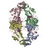
| ||||||||
|---|---|---|---|---|---|---|---|---|---|
| 1 |
| ||||||||
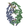
| Unit cell |
|
-Components
-Protein , 1 types, 4 molecules ABCD
| #1: Protein | Mass: 44140.449 Da / Num. of mol.: 4 / Mutation: G336V, C81A, C83A, C250A, C282A Source method: isolated from a genetically manipulated source Source: (gene. exp.) |
|---|
-Non-polymers , 5 types, 101 molecules 


| #2: Chemical | ChemComp-PO4 /  Mass: 94.971 Da / Num. of mol.: 8 / Source method: obtained synthetically / Formula: PO4 Mass: 94.971 Da / Num. of mol.: 8 / Source method: obtained synthetically / Formula: PO4#3: Chemical | ChemComp-NAI /  Mass: 665.441 Da / Num. of mol.: 4 / Source method: obtained synthetically / Formula: C21H29N7O14P2 Mass: 665.441 Da / Num. of mol.: 4 / Source method: obtained synthetically / Formula: C21H29N7O14P2#4: Chemical | ChemComp-CIT /  Mass: 192.124 Da / Num. of mol.: 6 / Source method: obtained synthetically / Formula: C6H8O7 Mass: 192.124 Da / Num. of mol.: 6 / Source method: obtained synthetically / Formula: C6H8O7#5: Chemical | ChemComp-SO4 / |  Mass: 96.063 Da / Num. of mol.: 1 / Source method: obtained synthetically / Formula: SO4 Mass: 96.063 Da / Num. of mol.: 1 / Source method: obtained synthetically / Formula: SO4#6: Water | ChemComp-HOH / | Mass: 18.015 Da / Num. of mol.: 82 / Source method: isolated from a natural source / Formula: H2O |
|---|
-Experimental details
-Experiment
| Experiment | Method: X-RAY DIFFRACTION / Number of used crystals: 1 |
|---|
- Sample preparation
| Crystal | Density Matthews: 2.9 Å3/Da / Density % sol: 57.6 % |
|---|---|
| Crystal grow | Temperature: 291 K / Method: vapor diffusion, hanging drop / pH: 5.5 Details: 1.5M AmSO4, 0.1M sodium citrate, pH 5.5, VAPOR DIFFUSION, HANGING DROP, temperature 291K |
-Data collection
| Diffraction | Mean temperature: 100 K |
|---|---|
| Diffraction source | Source: SYNCHROTRON / Site: APS  / Beamline: 19-ID / Wavelength: 0.9794 Å / Beamline: 19-ID / Wavelength: 0.9794 Å |
| Detector | Type: ADSC QUANTUM 315 / Detector: CCD / Date: Mar 16, 2005 / Details: Monochromator |
| Radiation | Monochromator: Si 111 / Protocol: SINGLE WAVELENGTH / Monochromatic (M) / Laue (L): M / Scattering type: x-ray |
| Radiation wavelength | Wavelength: 0.9794 Å / Relative weight: 1 |
| Reflection | Resolution: 2.6→50 Å / Num. obs: 61423 / % possible obs: 99.5 % / Redundancy: 18.7 % / Rmerge(I) obs: 0.165 / Net I/σ(I): 19.3 |
| Reflection shell | Resolution: 2.6→2.69 Å / Redundancy: 10.8 % / Rmerge(I) obs: 0.846 / Mean I/σ(I) obs: 1.64 / % possible all: 96.1 |
- Processing
| Software |
| ||||||||||||||||||||||||||||||||||||||||||||||||||||||||||||||||||||||||||||||||||||||||||
|---|---|---|---|---|---|---|---|---|---|---|---|---|---|---|---|---|---|---|---|---|---|---|---|---|---|---|---|---|---|---|---|---|---|---|---|---|---|---|---|---|---|---|---|---|---|---|---|---|---|---|---|---|---|---|---|---|---|---|---|---|---|---|---|---|---|---|---|---|---|---|---|---|---|---|---|---|---|---|---|---|---|---|---|---|---|---|---|---|---|---|---|
| Refinement | Method to determine structure: MOLECULAR REPLACEMENT Starting model: PDB entry 2PA3 Resolution: 2.6→35 Å / Cor.coef. Fo:Fc: 0.929 / Cor.coef. Fo:Fc free: 0.87 / SU B: 13.507 / SU ML: 0.286 / Cross valid method: THROUGHOUT / ESU R: 0.705 / ESU R Free: 0.345 / Stereochemistry target values: MAXIMUM LIKELIHOOD
| ||||||||||||||||||||||||||||||||||||||||||||||||||||||||||||||||||||||||||||||||||||||||||
| Solvent computation | Ion probe radii: 0.8 Å / Shrinkage radii: 0.8 Å / VDW probe radii: 1.4 Å / Solvent model: BABINET MODEL WITH MASK | ||||||||||||||||||||||||||||||||||||||||||||||||||||||||||||||||||||||||||||||||||||||||||
| Displacement parameters | Biso mean: 40.904 Å2
| ||||||||||||||||||||||||||||||||||||||||||||||||||||||||||||||||||||||||||||||||||||||||||
| Refinement step | Cycle: LAST / Resolution: 2.6→35 Å
| ||||||||||||||||||||||||||||||||||||||||||||||||||||||||||||||||||||||||||||||||||||||||||
| Refine LS restraints |
| ||||||||||||||||||||||||||||||||||||||||||||||||||||||||||||||||||||||||||||||||||||||||||
| LS refinement shell | Resolution: 2.6→2.667 Å / Total num. of bins used: 20
|
