[English] 日本語
Yorodumi
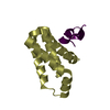
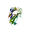
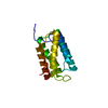
- PDB-2p8v: Crystal structure of human Homer3 EVH1 domain -

+
Open data


ID or keywords:

Loading...

-
Basic information

Entry
Database: PDB / ID: 2p8v
TitleCrystal structure of human Homer3 EVH1 domain
ComponentsHomer protein homolog 3
KeywordsSIGNALING PROTEIN / Homer3 / EVH1 domain
Function / homology
Function and homology information


G protein-coupled glutamate receptor binding / regulation of store-operated calcium entry / G protein-coupled glutamate receptor signaling pathway / basal part of cell / Neurexins and neuroligins / negative regulation of calcineurin-NFAT signaling cascade / negative regulation of interleukin-2 production / protein targeting / postsynaptic density / protein domain specific binding ...G protein-coupled glutamate receptor binding / regulation of store-operated calcium entry / G protein-coupled glutamate receptor signaling pathway / basal part of cell / Neurexins and neuroligins / negative regulation of calcineurin-NFAT signaling cascade / negative regulation of interleukin-2 production / protein targeting / postsynaptic density / protein domain specific binding / glutamatergic synapse / dendrite / identical protein binding / plasma membrane / cytosol / cytoplasm
Similarity search - Function
Homer, EVH1 domain / Homer family / WH1/EVH1 domain / WH1 domain / WH1 domain profile. / WASP homology region 1 / Pleckstrin-homology domain (PH domain)/Phosphotyrosine-binding domain (PTB) / PH-domain like / PH-like domain superfamily / Roll / Mainly Beta
Similarity search - Domain/homology
Homer protein homolog 3
Similarity search - Component
Biological speciesHomo sapiens (human)
MethodX-RAY DIFFRACTION / MOLECULAR REPLACEMENT / Resolution: 1.85 Å
AuthorsBouyain, S. / Tu, J. / Huang, G.N. / Worley, P.F. / Leahy, D.
CitationJournal: Science / Year: 2008
Title: NFAT binding and regulation of T cell activation by the cytoplasmic scaffolding Homer proteins.
Authors: Huang, G.N. / Huso, D.L. / Bouyain, S. / Tu, J. / McCorkell, K.A. / May, M.J. / Zhu, Y. / Lutz, M. / Collins, S. / Dehoff, M. / Kang, S. / Whartenby, K. / Powell, J. / Leahy, D. / Worley, P.F.
History
DepositionMar 23, 2007Deposition site: RCSB / Processing site: RCSB
Revision 1.0Nov 20, 2007Provider: repository / Type: Initial release
Revision 1.1Jul 13, 2011Group: Version format compliance
Revision 1.2Oct 18, 2017Group: Refinement description / Category: software
Revision 1.3Aug 30, 2023Group: Data collection / Database references ...Data collection / Database references / Derived calculations / Refinement description
Category: chem_comp_atom / chem_comp_bond ...chem_comp_atom / chem_comp_bond / database_2 / pdbx_initial_refinement_model / struct_site
Item: _database_2.pdbx_DOI / _database_2.pdbx_database_accession ..._database_2.pdbx_DOI / _database_2.pdbx_database_accession / _struct_site.pdbx_auth_asym_id / _struct_site.pdbx_auth_comp_id / _struct_site.pdbx_auth_seq_id

-
Structure visualization

Structure viewerMolecule:
MolmilJmol/JSmol

Downloads & links

-
Assembly

Deposited unit
A: Homer protein homolog 3
hetero molecules


Theoretical massNumber of molelcules
Total (without water)13,2712
Polymers13,1751
Non-polymers961
Water1,33374
1


  • Idetical with deposited unit
  • defined by author
TypeNameSymmetry operationNumber
identity operation1_555x,y,z1
Unit cell
Length a, b, c (Å)49.470, 49.470, 80.710
Angle α, β, γ (deg.)90.000, 90.000, 120.000
Int Tables number154
Space group name H-MP3221

-
Components

#1: Protein Homer protein homolog 3 / / Homer-3


Mass: 13174.810 Da / Num. of mol.: 1
Source method: isolated from a genetically manipulated source
Source: (gene. exp.) Homo sapiens (human) / Gene: HOMER3 / Plasmid: pT7HMT / Production host: Escherichia coli (E. coli) / Strain (production host): B834(DE3) / References: UniProt: Q9NSC5
#2: Chemical ChemComp-SO4 / SULFATE ION / Sulfate


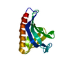
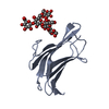
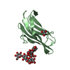
Mass: 96.063 Da / Num. of mol.: 1 / Source method: obtained synthetically / Formula: SO4
#3: Water ChemComp-HOH / water / Water


Mass: 18.015 Da / Num. of mol.: 74 / Source method: isolated from a natural source / Formula: H2O

-
Experimental details

-
Experiment

ExperimentMethod: X-RAY DIFFRACTION / Number of used crystals: 1

-
Sample preparation

CrystalDensity Matthews: 2.16 Å3/Da / Density % sol: 43.14 %
Crystal growTemperature: 293 K / Method: vapor diffusion, hanging drop / pH: 7.4
Details: 0.2M HEPES pH 7.0-7.4, 30% PEG3350, 105 mM ammonium sulfate, VAPOR DIFFUSION, HANGING DROP, temperature 293K

-
Data collection

DiffractionMean temperature: 100 K
Diffraction sourceSource: ROTATING ANODE / Type: RIGAKU / Wavelength: 1.5418 Å
DetectorType: ADSC QUANTUM 4 / Detector: CCD / Date: Sep 13, 2003
RadiationProtocol: SINGLE WAVELENGTH / Monochromatic (M) / Laue (L): M / Scattering type: x-ray
Radiation wavelengthWavelength: 1.5418 Å / Relative weight: 1
ReflectionResolution: 1.85→30 Å / Num. obs: 10075 / % possible obs: 98.8 % / Rmerge(I) obs: 0.078 / Χ2: 1.01 / Net I/σ(I): 20.3
Reflection shellResolution: 1.85→1.92 Å / Rmerge(I) obs: 0.493 / Num. unique all: 930 / Χ2: 1.082 / % possible all: 94

-
Phasing

Phasing MRRfactor: 0.42 / Cor.coef. Fo:Fc: 0.601
Highest resolutionLowest resolution
Rotation3 Å29.35 Å
Translation3 Å29.35 Å

-
Processing

Software
NameVersionClassificationNB
DENZOdata reduction
SCALEPACKdata scaling
MOLREPphasing
CNSrefinement
PDB_EXTRACT2data extraction
ADSCQuantumdata collection
RefinementMethod to determine structure: MOLECULAR REPLACEMENT
Starting model: 1DDW
Resolution: 1.85→30 Å / σ(F): 0
RfactorNum. reflection% reflection
Rfree0.264 1018 10 %
Rwork0.222 --
obs-9808 96 %
Solvent computationBsol: 38.076 Å2
Displacement parametersBiso mean: 26.706 Å2
Baniso -1Baniso -2Baniso -3
1--0.09 Å2-0.703 Å20 Å2
2---0.09 Å20 Å2
3---0.18 Å2
Refinement stepCycle: LAST / Resolution: 1.85→30 Å
ProteinNucleic acidLigandSolventTotal
Num. atoms940 0 5 74 1019
Refine LS restraints
Refine-IDTypeDev idealDev ideal target
X-RAY DIFFRACTIONc_bond_d0.006
X-RAY DIFFRACTIONc_angle_d1.311
X-RAY DIFFRACTIONc_mcbond_it2.6141.5
X-RAY DIFFRACTIONc_scbond_it4.032
X-RAY DIFFRACTIONc_mcangle_it3.62
X-RAY DIFFRACTIONc_scangle_it5.5792.5
Xplor file
Refine-IDSerial noParam fileTopol file
X-RAY DIFFRACTION1protein_rep.paramprotein.top
X-RAY DIFFRACTION2dna-rna_rep.paramdna-rna.top
X-RAY DIFFRACTION3water_rep.paramwater.top
X-RAY DIFFRACTION4ion.paramion.top
X-RAY DIFFRACTION5sulf.paramsulf.top

+
About Yorodumi

-
News

-
Feb 9, 2022. New format data for meta-information of EMDB entries

New format data for meta-information of EMDB entries

  • Version 3 of the EMDB header file is now the official format.
  • The previous official version 1.9 will be removed from the archive.

Related info.:EMDB header

External links:wwPDB to switch to version 3 of the EMDB data model

-
Aug 12, 2020. Covid-19 info

Covid-19 info

URL: https://pdbj.org/emnavi/covid19.php

New page: Covid-19 featured information page in EM Navigator.

Related info.:Covid-19 info / Mar 5, 2020. Novel coronavirus structure data

+
Mar 5, 2020. Novel coronavirus structure data

Novel coronavirus structure data

Related info.:Yorodumi Speices / Aug 12, 2020. Covid-19 info

External links:COVID-19 featured content - PDBj / Molecule of the Month (242):Coronavirus Proteases

+
Jan 31, 2019. EMDB accession codes are about to change! (news from PDBe EMDB page)

EMDB accession codes are about to change! (news from PDBe EMDB page)

  • The allocation of 4 digits for EMDB accession codes will soon come to an end. Whilst these codes will remain in use, new EMDB accession codes will include an additional digit and will expand incrementally as the available range of codes is exhausted. The current 4-digit format prefixed with “EMD-” (i.e. EMD-XXXX) will advance to a 5-digit format (i.e. EMD-XXXXX), and so on. It is currently estimated that the 4-digit codes will be depleted around Spring 2019, at which point the 5-digit format will come into force.
  • The EM Navigator/Yorodumi systems omit the EMD- prefix.

Related info.:Q: What is EMD? / ID/Accession-code notation in Yorodumi/EM Navigator

External links:EMDB Accession Codes are Changing Soon! / Contact to PDBj

+
Jul 12, 2017. Major update of PDB

Major update of PDB

  • wwPDB released updated PDB data conforming to the new PDBx/mmCIF dictionary.
  • This is a major update changing the version number from 4 to 5, and with Remediation, in which all the entries are updated.
  • In this update, many items about electron microscopy experimental information are reorganized (e.g. em_software).
  • Now, EM Navigator and Yorodumi are based on the updated data.

External links:wwPDB Remediation / Enriched Model Files Conforming to OneDep Data Standards Now Available in the PDB FTP Archive

-
Yorodumi

Thousand views of thousand structures

  • Yorodumi is a browser for structure data from EMDB, PDB, SASBDB, etc.
  • This page is also the successor to EM Navigator detail page, and also detail information page/front-end page for Omokage search.
  • The word "yorodu" (or yorozu) is an old Japanese word meaning "ten thousand". "mi" (miru) is to see.

Related info.:EMDB / PDB / SASBDB / Comparison of 3 databanks / Yorodumi Search / Aug 31, 2016. New EM Navigator & Yorodumi / Yorodumi Papers / Jmol/JSmol / Function and homology information / Changes in new EM Navigator and Yorodumi

Read more