- PDB-2p7n: Crystal structure of the Pathogenicity island 1 effector protein ... -
+
Open data
ID or keywords:
Loading...
-
Basic information
Entry
Database: PDB / ID: 2p7n
Title
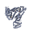
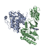
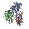
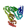
Crystal structure of the Pathogenicity island 1 effector protein from Chromobacterium violaceum. Northeast Structural Genomics Consortium (NESGC) target CvR69.
Components
Pathogenicity island 1 effector protein
Keywords
CELL INVASION / cvr69 / Pathogenicity island 1 effector protein / Structural Genomics / PSI-2 / Protein Structure Initiative / Northeast Structural Genomics Consortium / NESG
Function / homology
IpaD-like / IpaD-like / Type III secretion systems tip complex components / BipD-like superfamily / Type III secretion systems tip complex components / Up-down Bundle / extracellular region / Mainly Alpha / Translocator protein BipD
Function and homology information
Biological species
Chromobacterium violaceum (bacteria)
Method
X-RAY DIFFRACTION / SYNCHROTRON / MAD / Resolution: 2.8 Å
Method to determine structure: MAD / Resolution: 2.8→20 Å / FOM work R set: 0.779 / σ(F): 485 / Stereochemistry target values: Engh & Huber / Details: THE FRIEDEL PAIRS WERE USED FOR PHASING
Rfactor
Num. reflection
% reflection
Rfree
0.298
1496
8.8 %
Rwork
0.248
-
-
obs
-
15650
91.7 %
Solvent computation
Bsol: 11.085 Å2
Displacement parameters
Biso mean: 51.277 Å2
Baniso -1
Baniso -2
Baniso -3
1-
-6.022 Å2
0 Å2
0 Å2
2-
-
-19.066 Å2
0 Å2
3-
-
-
25.088 Å2
Refinement step
Cycle: LAST / Resolution: 2.8→20 Å
Protein
Nucleic acid
Ligand
Solvent
Total
Num. atoms
2514
0
0
15
2529
Refine LS restraints
Refine-ID
Type
Dev ideal
Dev ideal target
X-RAY DIFFRACTION
c_mcbond_it
1.542
1.5
X-RAY DIFFRACTION
c_scbond_it
2.451
2
X-RAY DIFFRACTION
c_mcangle_it
2.602
2
X-RAY DIFFRACTION
c_scangle_it
3.93
2.5
LS refinement shell
Refine-ID: X-RAY DIFFRACTION / Total num. of bins used: 29
Resolution (Å)
Rfactor Rfree
Num. reflection Rfree
Rfactor Rwork
Num. reflection Rwork
Num. reflection obs
2.8-2.83
0.5
31
0.379
385
416
2.83-2.87
0.382
63
0.411
439
502
2.87-2.9
0.436
37
0.372
430
467
2.9-2.94
0.474
43
0.389
426
469
2.94-2.98
0.403
54
0.321
442
496
2.98-3.02
0.283
37
0.322
471
508
3.02-3.07
0.316
52
0.324
487
539
3.07-3.12
0.532
54
0.35
450
504
3.12-3.17
0.427
43
0.304
480
523
3.17-3.22
0.338
43
0.259
500
543
3.22-3.28
0.337
62
0.302
450
512
3.28-3.34
0.253
43
0.254
527
570
3.34-3.41
0.312
55
0.3
470
525
3.41-3.49
0.325
48
0.26
527
575
3.49-3.57
0.272
48
0.243
498
546
3.57-3.65
0.307
48
0.227
449
497
3.65-3.75
0.304
49
0.245
455
504
3.75-3.86
0.315
56
0.218
484
540
3.86-3.99
0.24
50
0.214
523
573
3.99-4.13
0.269
57
0.209
529
586
4.13-4.29
0.295
55
0.188
505
560
4.29-4.48
0.252
45
0.228
555
600
4.48-4.72
0.31
73
0.201
491
564
4.72-5.01
0.239
68
0.225
529
597
5.01-5.39
0.278
48
0.245
534
582
5.39-5.92
0.259
68
0.273
526
594
5.92-6.75
0.37
53
0.241
542
595
6.75-8.39
0.293
60
0.241
518
578
8.39-20
0.213
53
0.206
532
585
Xplor file
Refine-ID
Serial no
Param file
X-RAY DIFFRACTION
1
protein_rep.par
X-RAY DIFFRACTION
2
water_rep.param
+
About Yorodumi
-
News
-
Feb 9, 2022. New format data for meta-information of EMDB entries
New format data for meta-information of EMDB entries
Version 3 of the EMDB header file is now the official format.
The previous official version 1.9 will be removed from the archive.
In the structure databanks used in Yorodumi, some data are registered as the other names, "COVID-19 virus" and "2019-nCoV". Here are the details of the virus and the list of structure data.
Jan 31, 2019. EMDB accession codes are about to change! (news from PDBe EMDB page)
EMDB accession codes are about to change! (news from PDBe EMDB page)
The allocation of 4 digits for EMDB accession codes will soon come to an end. Whilst these codes will remain in use, new EMDB accession codes will include an additional digit and will expand incrementally as the available range of codes is exhausted. The current 4-digit format prefixed with “EMD-” (i.e. EMD-XXXX) will advance to a 5-digit format (i.e. EMD-XXXXX), and so on. It is currently estimated that the 4-digit codes will be depleted around Spring 2019, at which point the 5-digit format will come into force.
The EM Navigator/Yorodumi systems omit the EMD- prefix.
Related info.:Q: What is EMD? / ID/Accession-code notation in Yorodumi/EM Navigator
Yorodumi is a browser for structure data from EMDB, PDB, SASBDB, etc.
This page is also the successor to EM Navigator detail page, and also detail information page/front-end page for Omokage search.
The word "yorodu" (or yorozu) is an old Japanese word meaning "ten thousand". "mi" (miru) is to see.
Related info.:EMDB / PDB / SASBDB / Comparison of 3 databanks / Yorodumi Search / Aug 31, 2016. New EM Navigator & Yorodumi / Yorodumi Papers / Jmol/JSmol / Function and homology information / Changes in new EM Navigator and Yorodumi
 Movie
Movie Controller

 Yorodumi
 Open data
 Basic information
 Components
 Keywords
 Function and homology information
 Chromobacterium violaceum (bacteria)
 X-RAY DIFFRACTION /
  Authors
 Citation
 Structure visualization
 Downloads & links
 Other downloads


 PDBj
PDBj Assembly
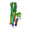

 Mass: 18.015 Da / Num. of mol.: 15 / Source method: isolated from a natural source / Formula: H2O
 Sample preparation
 / Beamline: X4A / Wavelength: 0.97912, 0.97897, 0.96862
 Processing