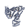
 Movie
Movie Controller


+ Open data

- Basic information
| Entry | Database: PDB / ID: 1n5u | ||||||
|---|---|---|---|---|---|---|---|
| Title | X-RAY STUDY OF HUMAN SERUM ALBUMIN COMPLEXED WITH HEME | ||||||
 Components Components | SERUM ALBUMIN | ||||||
 Keywords Keywords | PLASMA PROTEIN | ||||||
| Function / homology |  Function and homology information Function and homology informationbilirubin transport / Ciprofloxacin ADME / exogenous protein binding / cellular response to calcium ion starvation / enterobactin binding / Heme biosynthesis / HDL remodeling / negative regulation of mitochondrial depolarization / molecular carrier activity / Prednisone ADME ...bilirubin transport / Ciprofloxacin ADME / exogenous protein binding / cellular response to calcium ion starvation / enterobactin binding / Heme biosynthesis / HDL remodeling / negative regulation of mitochondrial depolarization / molecular carrier activity / Prednisone ADME / Heme degradation / Aspirin ADME / antioxidant activity / Scavenging of heme from plasma / toxic substance binding / Recycling of bile acids and salts / platelet alpha granule lumen / fatty acid binding / cellular response to starvation / Post-translational protein phosphorylation / Cytoprotection by HMOX1 / response to nutrient levels / Regulation of Insulin-like Growth Factor (IGF) transport and uptake by Insulin-like Growth Factor Binding Proteins (IGFBPs) / Platelet degranulation / pyridoxal phosphate binding / protein-folding chaperone binding / blood microparticle / endoplasmic reticulum lumen / copper ion binding / Golgi apparatus / endoplasmic reticulum / protein-containing complex / : / DNA binding / extracellular exosome / extracellular region / identical protein binding / nucleus Similarity search - Function | ||||||
| Biological species |  Homo sapiens (human) Homo sapiens (human) | ||||||
| Method |  X-RAY DIFFRACTION / SYNCHROTRON / MOLECULAR REPLACEMENT / Resolution: 1.9 Å X-RAY DIFFRACTION / SYNCHROTRON / MOLECULAR REPLACEMENT / Resolution: 1.9 Å | ||||||
 Authors Authors | Wardell, M. / Wang, Z. / Ho, J.X. / Robert, J. / Ruker, F. / Ruble, J. / Carter, D.C. | ||||||
 Citation Citation | Journal: Biochem.Biophys.Res.Commun. / Year: 2002 Title: The Atomic Structure of Human Methemalbumin at 1.9 A Authors: Wardell, M. / Wang, Z. / Ho, J.X. / Robert, J. / Ruker, F. / Ruble, J. / Carter, D.C. #1: Journal: Nature / Year: 1992Title: Atomic Structure and Chemistry of Human Serum Albumin Authors: He, X.M. / Carter, D.C. #2: Journal: Science / Year: 1990Title: Structure of Human Serum Albumin Authors: Carter, D.C. / He, X.M. #3: Journal: Science / Year: 1989Title: Three-Dimensional Structure of Human Serum Albumin Authors: Carter, D.C. / He, X.M. / Munson, S.H. / Twigg, P.D. / Gernert, K.M. / Broom, M.B. / Miller, T.Y. #4: Journal: Eur.J.Biochem. / Year: 1994Title: Preliminary Crystallographic Studies of Four Crystal Forms of Serum Albumin Authors: Carter, D.C. / Chang, B. / Ho, J.X. / Keeling, K. / Krishnasami, Z. | ||||||
| History |
|
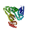
- Structure visualization
| Structure viewer | Molecule: MolmilJmol/JSmol |
|---|
- Downloads & links
-Download
| PDBx/mmCIF format | 1n5u.cif.gz | 140.5 KB | Display | PDBx/mmCIF format |
|---|---|---|---|---|
| PDB format | pdb1n5u.ent.gz | 108.6 KB | Display | PDB format |
| PDBx/mmJSON format | 1n5u.json.gz | Tree view | PDBx/mmJSON format | |
| Others |  Other downloads Other downloads |
-Validation report
| Arichive directory | https://data.pdbj.org/pub/pdb/validation_reports/n5/1n5uftp://data.pdbj.org/pub/pdb/validation_reports/n5/1n5u | HTTPS FTP |
|---|
-Related structure data
| Similar structure data |
|---|


-Links
 PDBj
PDBj


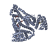
- Assembly
| Deposited unit | 
| ||||||||
|---|---|---|---|---|---|---|---|---|---|
| 1 |
| ||||||||
| Unit cell |
|
-Components
| #1: Protein | Mass: 66571.219 Da / Num. of mol.: 1 / Source method: isolated from a natural source / Source: (natural) Homo sapiens (human) / References: UniProt: P02768 | ||||||
|---|---|---|---|---|---|---|---|
| #2: Chemical | ChemComp-MYR /   Mass: 228.371 Da / Num. of mol.: 5 / Source method: obtained synthetically / Formula: C14H28O2 Mass: 228.371 Da / Num. of mol.: 5 / Source method: obtained synthetically / Formula: C14H28O2#3: Chemical | ChemComp-HEM / |   Mass: 616.487 Da / Num. of mol.: 1 / Source method: obtained synthetically / Formula: C34H32FeN4O4 Mass: 616.487 Da / Num. of mol.: 1 / Source method: obtained synthetically / Formula: C34H32FeN4O4#4: Water | ChemComp-HOH / |  Mass: 18.015 Da / Num. of mol.: 428 / Source method: isolated from a natural source / Formula: H2O Mass: 18.015 Da / Num. of mol.: 428 / Source method: isolated from a natural source / Formula: H2OHas protein modification | Y | |
-Experimental details
-Experiment
| Experiment | Method: X-RAY DIFFRACTION / Number of used crystals: 1 |
|---|
- Sample preparation
| Crystal | Density Matthews: 2.39 Å3/Da / Density % sol: 50.1 % |
|---|---|
| Crystal grow | Temperature: 293 K / Method: vapor diffusion, sitting drop / pH: 7.5 Details: PEG 3350, potassium phosphate, pH 7.5, VAPOR DIFFUSION, SITTING DROP, temperature 293K |
| Crystal grow | *PLUS Method: unknown / Details: Carter, D.C., (1994) Eur. J. Biochem., 226, 1049. |
-Data collection
| Diffraction | Mean temperature: 100 K | |||||||||
|---|---|---|---|---|---|---|---|---|---|---|
| Diffraction source | Source: SYNCHROTRON / Site: ALS  / Beamline: 5.0.3 / Wavelength: 1 / Wavelength: 1 Å / Beamline: 5.0.3 / Wavelength: 1 / Wavelength: 1 Å | |||||||||
| Detector | Type: ADSC QUANTUM 4 / Detector: CCD / Date: Aug 8, 2001 | |||||||||
| Radiation | Monochromator: SINGLE CRYSTAL, CYLINDRICALLY B / Protocol: SINGLE WAVELENGTH / Monochromatic (M) / Laue (L): M / Scattering type: x-ray | |||||||||
| Radiation wavelength |
| |||||||||
| Reflection | Resolution: 1.9→50 Å / Num. obs: 45420 / % possible obs: 90.3 % / Observed criterion σ(F): -3 / Redundancy: 2.89 % / Biso Wilson estimate: 18.1 Å2 / Rmerge(I) obs: 0.043 | |||||||||
| Reflection shell | Resolution: 1.9→1.97 Å / Redundancy: 2.11 % / Rmerge(I) obs: 0.298 / % possible all: 68.3 | |||||||||
| Reflection | *PLUS Highest resolution: 1.9 Å / % possible obs: 83.5 % / Redundancy: 2.6 % / Rmerge(I) obs: 0.037 | |||||||||
| Reflection shell | *PLUS Highest resolution: 1.9 Å / % possible obs: 56.3 % / Rmerge(I) obs: 0.266 |
- Processing
| Software |
| ||||||||||||||||||||||||||||||||||||
|---|---|---|---|---|---|---|---|---|---|---|---|---|---|---|---|---|---|---|---|---|---|---|---|---|---|---|---|---|---|---|---|---|---|---|---|---|---|
| Refinement | Method to determine structure: MOLECULAR REPLACEMENT / Resolution: 1.9→45.81 Å / Rfactor Rfree error: 0.006 / Isotropic thermal model: RESTRAINED / Cross valid method: THROUGHOUT / σ(F): 0 / Stereochemistry target values: Engh & Huber / Details: RESOLUTION-DEPENDENT WEIGHTING SCHEME
| ||||||||||||||||||||||||||||||||||||
| Solvent computation | Solvent model: FLAT MODEL / Bsol: 57.3643 Å2 / ksol: 0.375589 e/Å3 | ||||||||||||||||||||||||||||||||||||
| Displacement parameters | Biso mean: 29.4 Å2
| ||||||||||||||||||||||||||||||||||||
| Refine analyze |
| ||||||||||||||||||||||||||||||||||||
| Refinement step | Cycle: LAST / Resolution: 1.9→45.81 Å
| ||||||||||||||||||||||||||||||||||||
| Refine LS restraints |
| ||||||||||||||||||||||||||||||||||||
| LS refinement shell | Resolution: 1.9→2.02 Å / Rfactor Rfree error: 0.019 / Total num. of bins used: 6
| ||||||||||||||||||||||||||||||||||||
| Xplor file |
| ||||||||||||||||||||||||||||||||||||
| Refinement | *PLUS Highest resolution: 1.9 Å / Lowest resolution: 50 Å / % reflection Rfree: 5 % / Rfactor Rfree: 0.282 / Rfactor Rwork: 0.228 | ||||||||||||||||||||||||||||||||||||
| Solvent computation | *PLUS | ||||||||||||||||||||||||||||||||||||
| Displacement parameters | *PLUS | ||||||||||||||||||||||||||||||||||||
| Refine LS restraints | *PLUS
|
