[English] 日本語
Yorodumi
- PDB-2p7j: Crystal structure of the domain of putative sensory box/GGDEF fam... -

+
Open data


ID or keywords:

Loading...

-
Basic information

Entry
Database: PDB / ID: 2p7j
Title
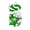
Crystal structure of the domain of putative sensory box/GGDEF family protein from Vibrio parahaemolyticus
ComponentsPutative sensory box/GGDEF family protein
KeywordsTRANSCRIPTION / Sensory box/GGDEF family protein / Structural genomics / MCSG / PSI-2 / Protein Structure Initiative / Midwest Center for Structural Genomics
Function / homology
Function and homology information


protein histidine kinase activity
Similarity search - Function
: / Bacterial histidine kinase, sensor domain / Periplasmic sensor-like domain superfamily / Diguanylate cyclase, GGDEF domain / diguanylate cyclase / Single alpha-helices involved in coiled-coils or other helix-helix interfaces - #170 / GGDEF domain profile. / GGDEF domain / PAS domain / PAS-associated, C-terminal ...: / Bacterial histidine kinase, sensor domain / Periplasmic sensor-like domain superfamily / Diguanylate cyclase, GGDEF domain / diguanylate cyclase / Single alpha-helices involved in coiled-coils or other helix-helix interfaces - #170 / GGDEF domain profile. / GGDEF domain / PAS domain / PAS-associated, C-terminal / PAC domain profile. / Nucleotide cyclase / PAS domain / Beta-Lactamase / PAS repeat profile. / PAS domain / Single alpha-helices involved in coiled-coils or other helix-helix interfaces / PAS domain superfamily / Reverse transcriptase/Diguanylate cyclase domain / Up-down Bundle / 2-Layer Sandwich / Mainly Alpha / Alpha Beta
Similarity search - Domain/homology
ACETIC ACID / Putative sensory box/GGDEF family protein
Similarity search - Component
Biological speciesVibrio parahaemolyticus RIMD 2210633 (bacteria)
MethodX-RAY DIFFRACTION / SYNCHROTRON / SAD / Resolution: 2.25 Å
AuthorsWu, R. / James, A. / Joachimiak, A. / Midwest Center for Structural Genomics (MCSG)
CitationJournal: To be Published
Title: The crystal structure of the domain of putative sensory box/GGDEF family protein from Vibrio parahaemolyticus
Authors: Wu, R. / James, A. / Joachimiak, A.
History
DepositionMar 20, 2007Deposition site: RCSB / Processing site: RCSB
Revision 1.0May 1, 2007Provider: repository / Type: Initial release
Revision 1.1May 1, 2008Group: Version format compliance
Revision 1.2Jul 13, 2011Group: Advisory / Version format compliance
Remark 300 BIOMOLECULE: 1 THIS ENTRY CONTAINS THE CRYSTALLOGRAPHIC ASYMMETRIC UNIT WHICH CONSISTS OF 2 ... BIOMOLECULE: 1 THIS ENTRY CONTAINS THE CRYSTALLOGRAPHIC ASYMMETRIC UNIT WHICH CONSISTS OF 2 CHAIN(S). AUTHORS STATE THAT THE BIOLOGICAL UNIT OF THIS PROTEIN IS A DIMER. SEE REMARK 350 FOR INFORMATION ON GENERATING THE BIOLOGICAL MOLECULE(S).

-
Structure visualization

Structure viewerMolecule:
MolmilJmol/JSmol

Downloads & links

-
Assembly

Deposited unit
A: Putative sensory box/GGDEF family protein
B: Putative sensory box/GGDEF family protein
hetero molecules


Theoretical massNumber of molelcules
Total (without water)66,8896
Polymers66,6142
Non-polymers2754
Water5,783321
1


  • Idetical with deposited unit
  • defined by author
TypeNameSymmetry operationNumber
identity operation1_555x,y,z1
Unit cell
Length a, b, c (Å)72.980, 81.596, 121.851
Angle α, β, γ (deg.)90.00, 90.00, 90.00
Int Tables number19
Space group name H-MP212121
DetailsThis protein exists as a dimer

-
Components

#1: Protein Putative sensory box/GGDEF family protein


Mass: 33306.918 Da / Num. of mol.: 2 / Fragment: Residues 27-312
Source method: isolated from a genetically manipulated source
Source: (gene. exp.) Vibrio parahaemolyticus RIMD 2210633 (bacteria)
Species: Vibrio parahaemolyticus / Strain: 223926 / Gene: VP0354 / Plasmid: PDM68 / Species (production host): Escherichia coli / Production host: Escherichia coli BL21 (bacteria) / Strain (production host): BL21 / References: UniProt: Q87SR8
#2: Chemical ChemComp-SO4 / SULFATE ION


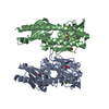
Mass: 96.063 Da / Num. of mol.: 2 / Source method: obtained synthetically / Formula: SO4
#3: Chemical ChemComp-NA / SODIUM ION


Mass: 22.990 Da / Num. of mol.: 1 / Source method: obtained synthetically / Formula: Na
#4: Chemical ChemComp-ACY / ACETIC ACID


Mass: 60.052 Da / Num. of mol.: 1 / Source method: obtained synthetically / Formula: C2H4O2
#5: Water ChemComp-HOH / water


Mass: 18.015 Da / Num. of mol.: 321 / Source method: isolated from a natural source / Formula: H2O

-
Experimental details

-
Experiment

ExperimentMethod: X-RAY DIFFRACTION / Number of used crystals: 1

-
Sample preparation

CrystalDensity Matthews: 2.73 Å3/Da / Density % sol: 54.97 %
Crystal growTemperature: 298 K / Method: vapor diffusion, sitting drop / pH: 7.5
Details: 0.2M LiSO4, 25% PEG 4000, pH 7.5, VAPOR DIFFUSION, SITTING DROP, temperature 298K

-
Data collection

DiffractionMean temperature: 100 K
Diffraction sourceSource: SYNCHROTRON / Site: APS / Beamline: 19-ID / Wavelength: 0.9793 Å
DetectorType: ADSC QUANTUM 315 / Detector: CCD / Date: Feb 4, 2007
RadiationMonochromator: Si 111 channel / Protocol: SINGLE WAVELENGTH / Monochromatic (M) / Laue (L): M / Scattering type: x-ray
Radiation wavelengthWavelength: 0.9793 Å / Relative weight: 1
ReflectionResolution: 2.25→50 Å / Num. all: 34928 / Num. obs: 33442 / % possible obs: 99.8 % / Observed criterion σ(I): 2 / Redundancy: 9 % / Biso Wilson estimate: 43 Å2 / Rmerge(I) obs: 0.11 / Net I/σ(I): 32.2
Reflection shellResolution: 2.25→2.33 Å / Redundancy: 8.8 % / Rmerge(I) obs: 0.583 / Mean I/σ(I) obs: 3.96 / Num. unique all: 3433 / % possible all: 100

-
Processing

Software
NameVersionClassification
REFMAC5.2.0019refinement
SBC-Collectdata collection
HKL-3000data reduction
HKL-3000data scaling
HKL-3000phasing
RefinementMethod to determine structure: SAD / Resolution: 2.25→38.69 Å / Cor.coef. Fo:Fc: 0.952 / Cor.coef. Fo:Fc free: 0.929 / SU B: 11.175 / SU ML: 0.149 / TLS residual ADP flag: LIKELY RESIDUAL / Cross valid method: THROUGHOUT / σ(F): 0 / σ(I): 0 / ESU R: 0.264 / ESU R Free: 0.21
Stereochemistry target values: MAXIMUM LIKELIHOOD WITH PHASES
Details: HYDROGENS HAVE BEEN ADDED IN THE RIDING POSITIONS
RfactorNum. reflection% reflectionSelection details
Rfree0.23863 1746 5 %RANDOM
Rwork0.19346 ---
all0.19577 33090 --
obs0.19577 33090 98.97 %-
Solvent computationIon probe radii: 0.8 Å / Shrinkage radii: 0.8 Å / VDW probe radii: 1.2 Å / Solvent model: MASK
Displacement parametersBiso mean: 42.187 Å2
Baniso -1Baniso -2Baniso -3
1-1.64 Å20 Å20 Å2
2--0.81 Å20 Å2
3----2.45 Å2
Refinement stepCycle: LAST / Resolution: 2.25→38.69 Å
ProteinNucleic acidLigandSolventTotal
Num. atoms4391 0 15 321 4727
Refine LS restraints
Refine-IDTypeDev idealDev ideal targetNumber
X-RAY DIFFRACTIONr_bond_refined_d0.0140.0224619
X-RAY DIFFRACTIONr_bond_other_d
X-RAY DIFFRACTIONr_angle_refined_deg1.5441.9636275
X-RAY DIFFRACTIONr_angle_other_deg
X-RAY DIFFRACTIONr_dihedral_angle_1_deg11.555562
X-RAY DIFFRACTIONr_dihedral_angle_2_deg37.48724.5240
X-RAY DIFFRACTIONr_dihedral_angle_3_deg14.41515813
X-RAY DIFFRACTIONr_dihedral_angle_4_deg19.2391531
X-RAY DIFFRACTIONr_chiral_restr0.1150.2674
X-RAY DIFFRACTIONr_gen_planes_refined0.0060.023581
X-RAY DIFFRACTIONr_gen_planes_other
X-RAY DIFFRACTIONr_nbd_refined0.2130.22048
X-RAY DIFFRACTIONr_nbd_other
X-RAY DIFFRACTIONr_nbtor_refined0.3060.23179
X-RAY DIFFRACTIONr_nbtor_other
X-RAY DIFFRACTIONr_xyhbond_nbd_refined0.1890.2353
X-RAY DIFFRACTIONr_xyhbond_nbd_other
X-RAY DIFFRACTIONr_metal_ion_refined
X-RAY DIFFRACTIONr_metal_ion_other
X-RAY DIFFRACTIONr_symmetry_vdw_refined0.2480.255
X-RAY DIFFRACTIONr_symmetry_vdw_other
X-RAY DIFFRACTIONr_symmetry_hbond_refined0.280.217
X-RAY DIFFRACTIONr_symmetry_hbond_other
X-RAY DIFFRACTIONr_symmetry_metal_ion_refined
X-RAY DIFFRACTIONr_symmetry_metal_ion_other
X-RAY DIFFRACTIONr_mcbond_it0.7091.52771
X-RAY DIFFRACTIONr_mcbond_other
X-RAY DIFFRACTIONr_mcangle_it1.37824502
X-RAY DIFFRACTIONr_scbond_it2.15431911
X-RAY DIFFRACTIONr_scangle_it3.4844.51773
X-RAY DIFFRACTIONr_rigid_bond_restr
X-RAY DIFFRACTIONr_sphericity_free
X-RAY DIFFRACTIONr_sphericity_bonded
LS refinement shellResolution: 2.25→2.31 Å / Total num. of bins used: 20
RfactorNum. reflection% reflection
Rfree0.257 110 -
Rwork0.209 2173 -
obs-2173 89.32 %
Refinement TLS params.Method: refined / Origin x: 43.3463 Å / Origin y: 28.0199 Å / Origin z: 9.8719 Å
111213212223313233
T-0.0815 Å2-0.0376 Å20.0194 Å2--0.0562 Å2-0.019 Å2---0.0792 Å2
L0.332 °2-0.0627 °20.1315 °2-0.5334 °2-0.1556 °2--0.6171 °2
S-0.0217 Å °0.0485 Å °-0.0119 Å °-0.0377 Å °0.0122 Å °-0.0057 Å °0.0506 Å °-0.0055 Å °0.0094 Å °
Refinement TLS group
IDRefine-IDRefine TLS-IDAuth asym-IDLabel asym-IDAuth seq-IDLabel seq-ID
1X-RAY DIFFRACTION1AA9 - 6010 - 61
2X-RAY DIFFRACTION1AA61 - 12062 - 121
3X-RAY DIFFRACTION1AA121 - 180122 - 181
4X-RAY DIFFRACTION1AA181 - 240182 - 241
5X-RAY DIFFRACTION1AA241 - 273242 - 274
6X-RAY DIFFRACTION1BB8 - 609 - 61
7X-RAY DIFFRACTION1BB61 - 12062 - 121
8X-RAY DIFFRACTION1BB121 - 180122 - 181
9X-RAY DIFFRACTION1BB181 - 240182 - 241
10X-RAY DIFFRACTION1BB241 - 281242 - 282

+
About Yorodumi

-
News

-
Feb 9, 2022. New format data for meta-information of EMDB entries

New format data for meta-information of EMDB entries

  • Version 3 of the EMDB header file is now the official format.
  • The previous official version 1.9 will be removed from the archive.

Related info.:EMDB header

External links:wwPDB to switch to version 3 of the EMDB data model

-
Aug 12, 2020. Covid-19 info

Covid-19 info

URL: https://pdbjlvh1.pdbj.org/emnavi/covid19.php

New page: Covid-19 featured information page in EM Navigator.

Related info.:Covid-19 info / Mar 5, 2020. Novel coronavirus structure data

+
Mar 5, 2020. Novel coronavirus structure data

Novel coronavirus structure data

Related info.:Yorodumi Speices / Aug 12, 2020. Covid-19 info

External links:COVID-19 featured content - PDBj / Molecule of the Month (242):Coronavirus Proteases

+
Jan 31, 2019. EMDB accession codes are about to change! (news from PDBe EMDB page)

EMDB accession codes are about to change! (news from PDBe EMDB page)

  • The allocation of 4 digits for EMDB accession codes will soon come to an end. Whilst these codes will remain in use, new EMDB accession codes will include an additional digit and will expand incrementally as the available range of codes is exhausted. The current 4-digit format prefixed with “EMD-” (i.e. EMD-XXXX) will advance to a 5-digit format (i.e. EMD-XXXXX), and so on. It is currently estimated that the 4-digit codes will be depleted around Spring 2019, at which point the 5-digit format will come into force.
  • The EM Navigator/Yorodumi systems omit the EMD- prefix.

Related info.:Q: What is EMD? / ID/Accession-code notation in Yorodumi/EM Navigator

External links:EMDB Accession Codes are Changing Soon! / Contact to PDBj

+
Jul 12, 2017. Major update of PDB

Major update of PDB

  • wwPDB released updated PDB data conforming to the new PDBx/mmCIF dictionary.
  • This is a major update changing the version number from 4 to 5, and with Remediation, in which all the entries are updated.
  • In this update, many items about electron microscopy experimental information are reorganized (e.g. em_software).
  • Now, EM Navigator and Yorodumi are based on the updated data.

External links:wwPDB Remediation / Enriched Model Files Conforming to OneDep Data Standards Now Available in the PDB FTP Archive

-
Yorodumi

Thousand views of thousand structures

  • Yorodumi is a browser for structure data from EMDB, PDB, SASBDB, etc.
  • This page is also the successor to EM Navigator detail page, and also detail information page/front-end page for Omokage search.
  • The word "yorodu" (or yorozu) is an old Japanese word meaning "ten thousand". "mi" (miru) is to see.

Related info.:EMDB / PDB / SASBDB / Comparison of 3 databanks / Yorodumi Search / Aug 31, 2016. New EM Navigator & Yorodumi / Yorodumi Papers / Jmol/JSmol / Function and homology information / Changes in new EM Navigator and Yorodumi

Read more