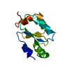
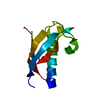
[English] 日本語
Yorodumi
- PDB-2p64: D domain of b-TrCP -

+
Open data


ID or keywords:

Loading...

-
Basic information

Entry
Database: PDB / ID: 2p64
TitleD domain of b-TrCP
ComponentsF-box/WD repeat protein 1A
KeywordsLIGASE / right handed super-helical bundle
Function / homology
Function and homology information


protein phosphorylated amino acid binding / ubiquitin ligase activator activity / positive regulation of ubiquitin protein ligase activity / positive regulation of circadian rhythm / regulation of canonical Wnt signaling pathway / branching involved in mammary gland duct morphogenesis / regulation of canonical NF-kappaB signal transduction / mammary gland epithelial cell proliferation / SCF ubiquitin ligase complex / SCF-dependent proteasomal ubiquitin-dependent protein catabolic process ...protein phosphorylated amino acid binding / ubiquitin ligase activator activity / positive regulation of ubiquitin protein ligase activity / positive regulation of circadian rhythm / regulation of canonical Wnt signaling pathway / branching involved in mammary gland duct morphogenesis / regulation of canonical NF-kappaB signal transduction / mammary gland epithelial cell proliferation / SCF ubiquitin ligase complex / SCF-dependent proteasomal ubiquitin-dependent protein catabolic process / Prolactin receptor signaling / non-canonical NF-kappaB signal transduction / ligase activity / positive regulation of proteolysis / cellular response to organic cyclic compound / ubiquitin-like ligase-substrate adaptor activity / protein K48-linked ubiquitination / negative regulation of smoothened signaling pathway / regulation of proteasomal protein catabolic process / MAP3K8 (TPL2)-dependent MAPK1/3 activation / protein dephosphorylation / SCF-beta-TrCP mediated degradation of Emi1 / NIK-->noncanonical NF-kB signaling / Vpu mediated degradation of CD4 / Deactivation of the beta-catenin transactivating complex / Dectin-1 mediated noncanonical NF-kB signaling / Degradation of GLI1 by the proteasome / Activation of NF-kappaB in B cells / GSK3B and BTRC:CUL1-mediated-degradation of NFE2L2 / Degradation of GLI2 by the proteasome / GLI3 is processed to GLI3R by the proteasome / protein destabilization / Degradation of beta-catenin by the destruction complex / regulation of circadian rhythm / negative regulation of DNA-binding transcription factor activity / CLEC7A (Dectin-1) signaling / beta-catenin binding / Wnt signaling pathway / FCERI mediated NF-kB activation / Interleukin-1 signaling / protein polyubiquitination / Regulation of PLK1 Activity at G2/M Transition / ubiquitin protein ligase activity / rhythmic process / Circadian Clock / Antigen processing: Ubiquitination & Proteasome degradation / Downstream TCR signaling / Neddylation / ubiquitin-dependent protein catabolic process / proteasome-mediated ubiquitin-dependent protein catabolic process / protein dimerization activity / regulation of cell cycle / protein ubiquitination / negative regulation of DNA-templated transcription / positive regulation of DNA-templated transcription / signal transduction / nucleoplasm / cytoplasm / cytosol
Similarity search - Function
Single alpha-helices involved in coiled-coils or other helix-helix interfaces - #1840 / D domain of beta-TrCP / D domain of beta-TrCP / D domain of beta-TrCP / A Receptor for Ubiquitination Targets / F-box domain profile. / F-box-like domain superfamily / F-box-like / F-box domain / Single alpha-helices involved in coiled-coils or other helix-helix interfaces ...Single alpha-helices involved in coiled-coils or other helix-helix interfaces - #1840 / D domain of beta-TrCP / D domain of beta-TrCP / D domain of beta-TrCP / A Receptor for Ubiquitination Targets / F-box domain profile. / F-box-like domain superfamily / F-box-like / F-box domain / Single alpha-helices involved in coiled-coils or other helix-helix interfaces / Helix non-globular / Special / G-protein beta WD-40 repeat / WD40 repeat, conserved site / Trp-Asp (WD) repeats signature. / Trp-Asp (WD) repeats profile. / Trp-Asp (WD) repeats circular profile. / WD domain, G-beta repeat / WD40 repeats / WD40 repeat / WD40-repeat-containing domain superfamily / WD40/YVTN repeat-like-containing domain superfamily
Similarity search - Domain/homology
: / F-box/WD repeat-containing protein 1A
Similarity search - Component
Biological speciesHomo sapiens (human)
MethodX-RAY DIFFRACTION / SYNCHROTRON / SAD / Resolution: 2.5 Å
AuthorsNeculai, D. / Orlicky, S. / Ceccarelli, D.
CitationJournal: Cell(Cambridge,Mass.) / Year: 2007
Title: Suprafacial orientation of the SCFCdc4 dimer accommodates multiple geometries for substrate ubiquitination.
Authors: Tang, X. / Orlicky, S. / Lin, Z. / Willems, A. / Neculai, D. / Ceccarelli, D. / Mercurio, F. / Shilton, B.H. / Sicheri, F. / Tyers, M.
History
DepositionMar 16, 2007Deposition site: RCSB / Processing site: RCSB
Revision 1.0Jun 19, 2007Provider: repository / Type: Initial release
Revision 1.1May 1, 2008Group: Version format compliance
Revision 1.2Jul 13, 2011Group: Advisory / Version format compliance

-
Structure visualization

Structure viewerMolecule:
MolmilJmol/JSmol

Downloads & links

-
Assembly

Deposited unit
A: F-box/WD repeat protein 1A
B: F-box/WD repeat protein 1A
hetero molecules


Theoretical massNumber of molelcules
Total (without water)12,7563
Polymers12,6442
Non-polymers1121
Water19811
1


  • Idetical with deposited unit
  • defined by author&software
TypeNameSymmetry operationNumber
identity operation1_555x,y,z1
Buried area3800 Å2
ΔGint-37 kcal/mol
Surface area6230 Å2
MethodPISA
Unit cell
Length a, b, c (Å)71.316, 71.316, 116.301
Angle α, β, γ (deg.)90.00, 90.00, 90.00
Int Tables number96
Space group name H-MP43212

-
Components

#1: Protein F-box/WD repeat protein 1A / F-box and WD repeats protein beta-TrCP / E3RSIkappaB / pIkappaBalpha-E3 receptor subunit


Mass: 6321.773 Da / Num. of mol.: 2 / Fragment: D domain
Source method: isolated from a genetically manipulated source
Source: (gene. exp.) Homo sapiens (human) / Gene: BTRC, BTRCP, FBW1A, FBXW1A / Plasmid: pGEX / Species (production host): Escherichia coli / Production host: Escherichia coli BL21 (bacteria) / Strain (production host): BL21 / References: UniProt: Q9Y297
#2: Chemical ChemComp-CD / CADMIUM ION


Mass: 112.411 Da / Num. of mol.: 1 / Source method: obtained synthetically / Formula: Cd
#3: Water ChemComp-HOH / water


Mass: 18.015 Da / Num. of mol.: 11 / Source method: isolated from a natural source / Formula: H2O

-
Experimental details

-
Experiment

ExperimentMethod: X-RAY DIFFRACTION / Number of used crystals: 1

-
Sample preparation

Crystal growTemperature: 293 K / Method: vapor diffusion, hanging drop / pH: 7.5
Details: 200 mM MgCl2, 5 mM CdCl2, pH 7.5, VAPOR DIFFUSION, HANGING DROP, temperature 293K

-
Data collection

DiffractionMean temperature: 100 K
Diffraction sourceSource: SYNCHROTRON / Site: APS / Beamline: 8-BM / Wavelength: 0.97906 Å
DetectorType: ADSC QUANTUM 315 / Detector: CCD / Date: Nov 5, 2005 / Details: mirrors
RadiationMonochromator: double-crystal monochromator SI(111) / Protocol: SINGLE WAVELENGTH / Monochromatic (M) / Laue (L): M / Scattering type: x-ray
Radiation wavelengthWavelength: 0.97906 Å / Relative weight: 1
ReflectionResolution: 2.4→30 Å / Num. all: 12340 / Num. obs: 11999 / % possible obs: 97.2 % / Observed criterion σ(I): 3 / Rmerge(I) obs: 0.246 / Net I/σ(I): 18.8
Reflection shellResolution: 2.4→2.5 Å / Rmerge(I) obs: 0.246 / Mean I/σ(I) obs: 3.19 / Num. unique all: 1100 / % possible all: 82.4

-
Processing

Software
NameVersionClassification
REFMAC5.2.0019refinement
CBASSdata collection
HKL-2000data reduction
HKL-2000data scaling
SHELXDphasing
RefinementMethod to determine structure: SAD / Resolution: 2.5→19.78 Å / Cor.coef. Fo:Fc: 0.935 / Cor.coef. Fo:Fc free: 0.91 / SU B: 11.644 / SU ML: 0.129 / TLS residual ADP flag: LIKELY RESIDUAL / Cross valid method: THROUGHOUT / ESU R: 0.219 / ESU R Free: 0.199 / Stereochemistry target values: MAXIMUM LIKELIHOOD
RfactorNum. reflection% reflectionSelection details
Rfree0.26679 536 4.9 %RANDOM
Rwork0.23854 ---
obs0.23986 10345 100 %-
Solvent computationIon probe radii: 0.8 Å / Shrinkage radii: 0.8 Å / VDW probe radii: 1.4 Å / Solvent model: MASK
Displacement parametersBiso mean: 54.936 Å2
Baniso -1Baniso -2Baniso -3
1--0.05 Å20 Å20 Å2
2---0.05 Å20 Å2
3---0.1 Å2
Refinement stepCycle: LAST / Resolution: 2.5→19.78 Å
ProteinNucleic acidLigandSolventTotal
Num. atoms868 0 1 11 880
Refine LS restraints
Refine-IDTypeDev idealDev ideal targetNumber
X-RAY DIFFRACTIONr_bond_refined_d0.0160.021906
X-RAY DIFFRACTIONr_angle_refined_deg1.5441.9131215
X-RAY DIFFRACTIONr_dihedral_angle_1_deg4.9925103
X-RAY DIFFRACTIONr_dihedral_angle_2_deg43.34225.4951
X-RAY DIFFRACTIONr_dihedral_angle_3_deg19.9615163
X-RAY DIFFRACTIONr_chiral_restr0.1160.2118
X-RAY DIFFRACTIONr_gen_planes_refined0.0060.02698
X-RAY DIFFRACTIONr_nbd_refined0.2080.2398
X-RAY DIFFRACTIONr_nbtor_refined0.3020.2600
X-RAY DIFFRACTIONr_xyhbond_nbd_refined0.1560.216
X-RAY DIFFRACTIONr_symmetry_vdw_refined0.3310.225
X-RAY DIFFRACTIONr_symmetry_hbond_refined0.3020.24
X-RAY DIFFRACTIONr_symmetry_metal_ion_refined0.3590.22
X-RAY DIFFRACTIONr_mcbond_it1.2081.5536
X-RAY DIFFRACTIONr_mcangle_it1.5312830
X-RAY DIFFRACTIONr_scbond_it2.6963435
X-RAY DIFFRACTIONr_scangle_it3.4534.5385
LS refinement shellResolution: 2.5→2.564 Å / Total num. of bins used: 20
RfactorNum. reflection% reflection
Rfree0.356 40 -
Rwork0.303 728 -
obs--100 %
Refinement TLS params.

Method: refined / Refine-ID: X-RAY DIFFRACTION

IDL112)L122)L132)L222)L232)L332)S11 (Å °)S12 (Å °)S13 (Å °)S21 (Å °)S22 (Å °)S23 (Å °)S31 (Å °)S32 (Å °)S33 (Å °)T112)T122)T132)T222)T232)T332)Origin x (Å)Origin y (Å)Origin z (Å)
16.027-1.7441-0.90435.60781.85215.9959-0.0871-0.21420.12680.0150.22-0.54270.20510.4123-0.133-0.0845-0.0289-0.04990.0716-0.05360.038967.319744.075555.5159
24.4588-1.53511.01993.3536-0.78054.18150.08890.0462-0.4158-0.10710.07760.02040.18010.1254-0.16660.0325-0.0424-0.0779-0.0171-0.02470.03165.151742.830656.5596
Refinement TLS group
IDRefine-IDRefine TLS-IDAuth asym-IDLabel asym-IDAuth seq-IDLabel seq-ID
1X-RAY DIFFRACTION1AA127 - 1772 - 52
2X-RAY DIFFRACTION2BB127 - 1772 - 52

+
About Yorodumi

-
News

-
Feb 9, 2022. New format data for meta-information of EMDB entries

New format data for meta-information of EMDB entries

  • Version 3 of the EMDB header file is now the official format.
  • The previous official version 1.9 will be removed from the archive.

Related info.:EMDB header

External links:wwPDB to switch to version 3 of the EMDB data model

-
Aug 12, 2020. Covid-19 info

Covid-19 info

URL: https://pdbjlvh1.pdbj.org/emnavi/covid19.php

New page: Covid-19 featured information page in EM Navigator.

Related info.:Covid-19 info / Mar 5, 2020. Novel coronavirus structure data

+
Mar 5, 2020. Novel coronavirus structure data

Novel coronavirus structure data

Related info.:Yorodumi Speices / Aug 12, 2020. Covid-19 info

External links:COVID-19 featured content - PDBj / Molecule of the Month (242):Coronavirus Proteases

+
Jan 31, 2019. EMDB accession codes are about to change! (news from PDBe EMDB page)

EMDB accession codes are about to change! (news from PDBe EMDB page)

  • The allocation of 4 digits for EMDB accession codes will soon come to an end. Whilst these codes will remain in use, new EMDB accession codes will include an additional digit and will expand incrementally as the available range of codes is exhausted. The current 4-digit format prefixed with “EMD-” (i.e. EMD-XXXX) will advance to a 5-digit format (i.e. EMD-XXXXX), and so on. It is currently estimated that the 4-digit codes will be depleted around Spring 2019, at which point the 5-digit format will come into force.
  • The EM Navigator/Yorodumi systems omit the EMD- prefix.

Related info.:Q: What is EMD? / ID/Accession-code notation in Yorodumi/EM Navigator

External links:EMDB Accession Codes are Changing Soon! / Contact to PDBj

+
Jul 12, 2017. Major update of PDB

Major update of PDB

  • wwPDB released updated PDB data conforming to the new PDBx/mmCIF dictionary.
  • This is a major update changing the version number from 4 to 5, and with Remediation, in which all the entries are updated.
  • In this update, many items about electron microscopy experimental information are reorganized (e.g. em_software).
  • Now, EM Navigator and Yorodumi are based on the updated data.

External links:wwPDB Remediation / Enriched Model Files Conforming to OneDep Data Standards Now Available in the PDB FTP Archive

-
Yorodumi

Thousand views of thousand structures

  • Yorodumi is a browser for structure data from EMDB, PDB, SASBDB, etc.
  • This page is also the successor to EM Navigator detail page, and also detail information page/front-end page for Omokage search.
  • The word "yorodu" (or yorozu) is an old Japanese word meaning "ten thousand". "mi" (miru) is to see.

Related info.:EMDB / PDB / SASBDB / Comparison of 3 databanks / Yorodumi Search / Aug 31, 2016. New EM Navigator & Yorodumi / Yorodumi Papers / Jmol/JSmol / Function and homology information / Changes in new EM Navigator and Yorodumi

Read more