Mass: 18.015 Da / Num. of mol.: 3 / Source method: isolated from a natural source / Formula: H2O
Has protein modification
Y
-
Experimental details
-
Experiment
Experiment
Method: X-RAY DIFFRACTION / Number of used crystals: 1
-
Sample preparation
Crystal
Density Matthews: 3.58 Å3/Da / Density % sol: 65.61 %
Crystal grow
Temperature: 294 K / Method: vapor diffusion, sitting drop / pH: 8.5 Details: 2.5 M Ammonium sulfate, 0.1 M Tris buffer, pH 8.5, VAPOR DIFFUSION, SITTING DROP, temperature 294K
Resolution: 2.45→2.52 Å / Redundancy: 10.4 % / Rmerge(I) obs: 0.852 / Mean I/σ(I) obs: 2.49 / Num. unique all: 541 / Χ2: 0.936 / % possible all: 100
-
Processing
Software
Name
Version
Classification
NB
PHENIX
refinement
SCALEPACK
datascaling
REFMAC
refinement
PDB_EXTRACT
3.005
dataextraction
SBC-Collect
datacollection
HKL-3000
datareduction
HKL-3000
datascaling
PHENIX
phasing
DENZO
datareduction
Refinement
Method to determine structure: SAD / Resolution: 2.46→32.1 Å / σ(F): 0 / σ(I): 0 / Stereochemistry target values: Engh & Huber Details: ANALYSIS OF THE DIFFRACTION DATA INDICATED HEMIHEDERAL TWINNING WITH A TWIN FRACTION OF 0.317 AND WITH A TWIN LAW OF K,H,-L.
Rfactor
Num. reflection
% reflection
Selection details
Rfree
0.2475
299
-
Random
Rwork
0.1935
-
-
-
all
-
6097
-
-
obs
-
6097
98.11 %
-
Displacement parameters
Biso mean: 73.624 Å2
Refinement step
Cycle: LAST / Resolution: 2.46→32.1 Å
Protein
Nucleic acid
Ligand
Solvent
Total
Num. atoms
721
0
5
3
729
Refine LS restraints
Refine-ID
Type
Dev ideal
X-RAY DIFFRACTION
f_bond_d
0.008
X-RAY DIFFRACTION
f_angle_deg
1.308
+
About Yorodumi
-
News
-
Feb 9, 2022. New format data for meta-information of EMDB entries
New format data for meta-information of EMDB entries
Version 3 of the EMDB header file is now the official format.
The previous official version 1.9 will be removed from the archive.
In the structure databanks used in Yorodumi, some data are registered as the other names, "COVID-19 virus" and "2019-nCoV". Here are the details of the virus and the list of structure data.
Jan 31, 2019. EMDB accession codes are about to change! (news from PDBe EMDB page)
EMDB accession codes are about to change! (news from PDBe EMDB page)
The allocation of 4 digits for EMDB accession codes will soon come to an end. Whilst these codes will remain in use, new EMDB accession codes will include an additional digit and will expand incrementally as the available range of codes is exhausted. The current 4-digit format prefixed with “EMD-” (i.e. EMD-XXXX) will advance to a 5-digit format (i.e. EMD-XXXXX), and so on. It is currently estimated that the 4-digit codes will be depleted around Spring 2019, at which point the 5-digit format will come into force.
The EM Navigator/Yorodumi systems omit the EMD- prefix.
Related info.:Q: What is EMD? / ID/Accession-code notation in Yorodumi/EM Navigator
Yorodumi is a browser for structure data from EMDB, PDB, SASBDB, etc.
This page is also the successor to EM Navigator detail page, and also detail information page/front-end page for Omokage search.
The word "yorodu" (or yorozu) is an old Japanese word meaning "ten thousand". "mi" (miru) is to see.
Related info.:EMDB / PDB / SASBDB / Comparison of 3 databanks / Yorodumi Search / Aug 31, 2016. New EM Navigator & Yorodumi / Yorodumi Papers / Jmol/JSmol / Function and homology information / Changes in new EM Navigator and Yorodumi
 Movie
Movie Controller

 Yorodumi
 Open data
 Basic information
 Components
 Keywords
 Function and homology information
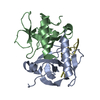
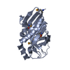
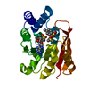
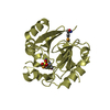
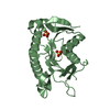
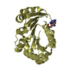
 Archaeoglobus fulgidus DSM 4304 (archaea)
 X-RAY DIFFRACTION /
  Authors
 Citation
 Structure visualization
 Downloads & links
 Other downloads


 PDBj
PDBj


 Assembly


 Mass: 96.063 Da / Num. of mol.: 1 / Source method: obtained synthetically / Formula: SO4
 Mass: 18.015 Da / Num. of mol.: 3 / Source method: isolated from a natural source / Formula: H2O
 Sample preparation
 / Beamline: 19-BM / Wavelength: 0.9792 Å
 Processing