Mass: 18.015 Da / Num. of mol.: 117 / Source method: isolated from a natural source / Formula: H2O
Has protein modification
Y
Nonpolymer details
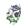
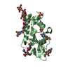
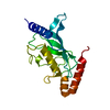
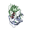
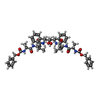
THE INHIBITOR IS A C2 SYMMETRIC HIV PROTEASE
-
Experimental details
-
Experiment
Experiment
Method: X-RAY DIFFRACTION / Number of used crystals: 1
-
Sample preparation
Crystal
Density Matthews: 2.05 Å3/Da / Density % sol: 39.99 %
Crystal grow
Temperature: 291 K / Method: vapor diffusion, hanging drop / pH: 6.2 Details: 0.8M ammonium sulfate, 0.1M sodium cacodylate. The crystals nucleated at 277K and were transfered to 291K for further growth. The protein to well proportion was 2:1, pH 6.2, VAPOR DIFFUSION, HANGING DROP
Resolution: 2.1→53.225 Å / Num. obs: 10162 / % possible obs: 99.7 % / Redundancy: 2.5 % / Rmerge(I) obs: 0.046 / Rsym value: 0.046 / Net I/σ(I): 10.8
Reflection shell
Diffraction-ID: 1,2
Resolution (Å)
Redundancy (%)
Rmerge(I) obs
Mean I/σ(I) obs
Num. measured all
Num. unique all
Rsym value
% possible all
2.1-2.21
2.4
0.318
2.1
3638
1491
0.318
99.8
2.21-2.35
2.4
0.234
2.8
3398
1389
0.234
99.8
2.35-2.51
2.4
0.177
2.6
3199
1307
0.177
100
2.51-2.71
2.4
0.114
5.9
3018
1234
0.114
100
2.71-2.97
2.5
0.072
9.6
2784
1131
0.072
100
2.97-3.32
2.5
0.047
13.7
2543
1028
0.047
100
3.32-3.83
2.5
0.034
17.6
2270
909
0.034
100
3.83-4.7
2.5
0.026
20.1
1915
762
0.026
99.7
4.7-6.64
2.5
0.023
24.4
1516
599
0.023
99.1
6.64-32.21
2.6
0.026
16.9
800
312
0.026
94.2
-
Processing
Software
Name
Version
Classification
NB
SCALA
datascaling
REFMAC
5.2.0003
refinement
PDB_EXTRACT
2
dataextraction
MAR345
345DTB
datacollection
MOSFLM
datareduction
CCP4
(SCALA)
datascaling
PHASER
phasing
Refinement
Method to determine structure: MOLECULAR REPLACEMENT Starting model: Crystal Structure of the multi-drug resistant mutant subtype B HIV protease complexed with TL-3 inhibitor Resolution: 2.1→25.27 Å / Cor.coef. Fo:Fc: 0.955 / Cor.coef. Fo:Fc free: 0.925 / SU B: 14.02 / SU ML: 0.202 / Cross valid method: THROUGHOUT / σ(F): 0 / ESU R Free: 0.262 / Stereochemistry target values: MAXIMUM LIKELIHOOD / Details: HYDROGENS HAVE BEEN ADDED IN THE RIDING POSITIONS
Rfactor
Num. reflection
% reflection
Selection details
Rfree
0.265
1008
10 %
RANDOM
Rwork
0.194
-
-
-
obs
0.201
10121
99.67 %
-
Solvent computation
Ion probe radii: 0.8 Å / Shrinkage radii: 0.8 Å / VDW probe radii: 1.2 Å / Solvent model: MASK
In the structure databanks used in Yorodumi, some data are registered as the other names, "COVID-19 virus" and "2019-nCoV". Here are the details of the virus and the list of structure data.
Jan 31, 2019. EMDB accession codes are about to change! (news from PDBe EMDB page)
EMDB accession codes are about to change! (news from PDBe EMDB page)
The allocation of 4 digits for EMDB accession codes will soon come to an end. Whilst these codes will remain in use, new EMDB accession codes will include an additional digit and will expand incrementally as the available range of codes is exhausted. The current 4-digit format prefixed with “EMD-” (i.e. EMD-XXXX) will advance to a 5-digit format (i.e. EMD-XXXXX), and so on. It is currently estimated that the 4-digit codes will be depleted around Spring 2019, at which point the 5-digit format will come into force.
The EM Navigator/Yorodumi systems omit the EMD- prefix.
Related info.:Q: What is EMD? / ID/Accession-code notation in Yorodumi/EM Navigator
Yorodumi is a browser for structure data from EMDB, PDB, SASBDB, etc.
This page is also the successor to EM Navigator detail page, and also detail information page/front-end page for Omokage search.
The word "yorodu" (or yorozu) is an old Japanese word meaning "ten thousand". "mi" (miru) is to see.
Related info.:EMDB / PDB / SASBDB / Comparison of 3 databanks / Yorodumi Search / Aug 31, 2016. New EM Navigator & Yorodumi / Yorodumi Papers / Jmol/JSmol / Function and homology information / Changes in new EM Navigator and Yorodumi
 Movie
Movie Controller

 Yorodumi
 Open data
 Basic information
 Components
 Keywords
 Function and homology information
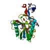
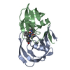
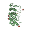
 Human immunodeficiency virus 1
 X-RAY DIFFRACTION /
  Authors
 Citation
 Structure visualization
 Downloads & links
 Other downloads


 PDBj
PDBj


 Assembly
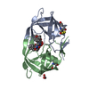

 Type: peptide-like, Peptide-like / Class: Inhibitor / Mass: 909.077 Da / Num. of mol.: 1 / Source method: obtained synthetically / Formula: C50H64N6O10
 Mass: 60.052 Da / Num. of mol.: 3 / Source method: obtained synthetically / Formula: C2H4O2
 Mass: 18.015 Da / Num. of mol.: 117 / Source method: isolated from a natural source / Formula: H2O
 Sample preparation
 / Beamline: D03B-MX1 / Wavelength: 1.43
  Processing